. 2017 Jun 20;8(25):41154-41165.
doi: 10.18632/oncotarget.17074.

The antigen-binding fragment of human gamma immunoglobulin prevents amyloid β-peptide folding into β-sheet to form oligomers

Affiliations

The antigen-binding fragment of human gamma immunoglobulin prevents amyloid β-peptide folding into β-sheet to form oligomers

Victòria Valls-Comamala et al. Oncotarget. .

Abstract

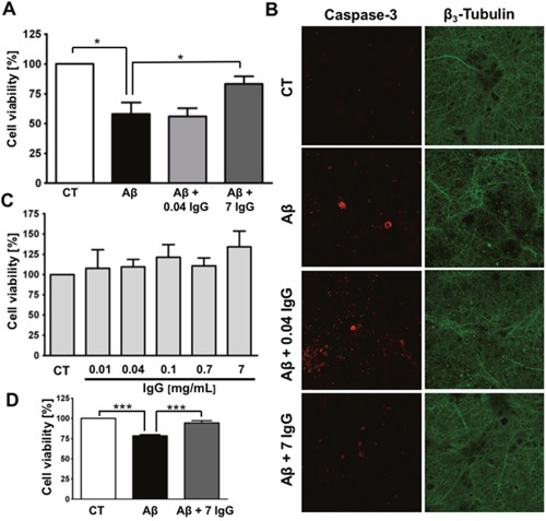
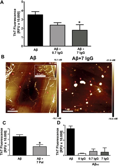
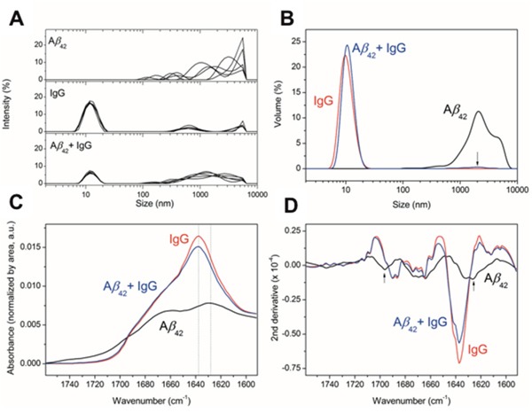
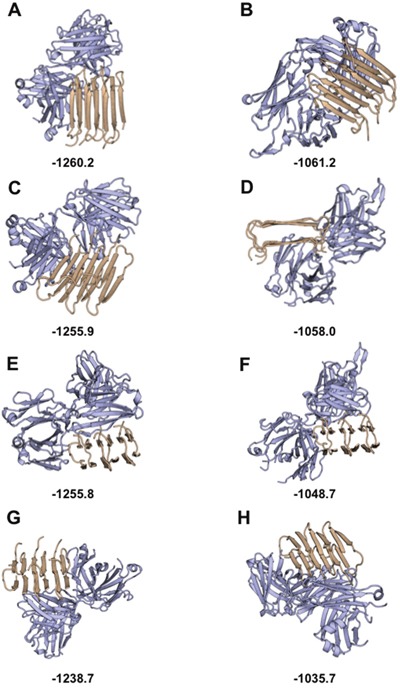
The amyloid beta-peptide (Aβ) plays a leading role in Alzheimer's disease (AD) physiopathology. Even though monomeric forms of Aβ are harmless to cells, Aβ can aggregate into β-sheet oligomers and fibrils, which are both neurotoxic. Therefore, one of the main therapeutic approaches to cure or delay AD onset and progression is targeting Aβ aggregation. In the present study, we show that a pool of human gamma immunoglobulins (IgG) protected cortical neurons from the challenge with Aβ oligomers, as assayed by MTT reduction, caspase-3 activation and cytoskeleton integrity. In addition, we report the inhibitory effect of IgG on Aβ aggregation, as shown by Thioflavin T assay, size exclusion chromatography and atomic force microscopy. Similar results were obtained with Palivizumab, a human anti-sincitial virus antibody. In order to dissect the important domains, we cleaved the pool of human IgG with papain to obtain Fab and Fc fragments. Using these cleaved fragments, we functionally identified Fab as the immunoglobulin fragment inhibiting Aβ aggregation, a result that was further confirmed by an in silico structural model. Interestingly, bioinformatic tools show a highly conserved structure able to bind amyloid in the Fab region. Overall, our data strongly support the inhibitory effect of human IgG on Aβ aggregation and its neuroprotective role.

Keywords: Alzheimer’s disease; Fab; amyloid; immunoglobulin; oligomers.


Conflict of interest statement

CONFLICTS OF INTEREST

The authors declare that they have no conflicts of interest.

Figures

Figure 1
Figure 1. IgG protects cortical neurons against amyloid toxicity
(A) Cell viability was assayed by MTT reduction in cortical neurons treated with 10 μM Aβ1-42 or 10 μM Aβ1-42 + IgG at either 0.04 or 7 mg/mL for 24 h. Data are the mean ± SEM of 6 independent experiments. *p < 0.05 by one-way ANOVA using Newman-Keuls post-test. (B) Immunofluorescence study of cortical neurons treated with 10 μM Aβ1-42, 10 μM Aβ1-42 + 0.04 mg/mL of IgG or 10 μM Aβ1-42 + 7 mg/mL of IgG for 24 h. Staining for active caspase-3 is shown in red and for β3-tubulin in green. β3-tubulin is an overlap of images of the same area (C) Cell viability was assayed in cortical neurons by MTT reduction treated with increasing concentrations of IgG for 24 h. Data are the mean ± SEM of 4 independent experiments. (D) Cell viability was assayed by MTT reduction in endothelial cells treated with 0.1 μM Aβ1-40 or 0.1 μM Aβ1-40 + IgG 7 mg/mL for 24 h. Data are the mean ± SEM of 5 independent experiments. *** p < 0.001 by one-way ANOVA using Newman-Keuls post-test.
Figure 2
Figure 2. IgG inhibits amyloid aggregation
(A) Aggregation assay of 27 μM Aβ1-42 with or without IgG at 0.7 and 7 mg/mL during 96 h. ThT fluorescence was measured at 96 h. Data are the mean ± SEM of 3 independent experiments. * p < 0.05 compared to control by one-way ANOVA using Newman-Keuls post-test. (B) AFM images of 88,6 μM Aβ1-42 (left) and 88,6 μM Aβ1-42 treated with 7 mg/mL of IgG (right) for 96 h. The inset in the left image shows a cluster of oligomers. Arrows indicate mature fibers (left) and amorphous globular structures (right) (C) Aggregation assay of 27 μM Aβ1-42 with or without Palivizumab at 7 mg/mL during 96 h. ThT fluorescence was measured at 96 h. Data are the mean ± SEM of 3 independent experiments. * p < 0.05 by t-student test. (D) Aggregation assay of 27 μM Aβ1-42, 27 μM Aβ1-42 scramble (Aβsc) and 27 μM Aβsc co-incubated with IgG at 0.7 mg/mL and 7 mg/mL. ThT fluorescence was measured at 96 h. Data are the mean ± SEM of 3 independent experiments.
Figure 3
Figure 3. Effect of IgG on Aβ oligomerization
(A) DLS. Size distribution measured by DLS as a function of signal intensity of particles for 5 DLS measurements of 10 scans each. (B) Size distribution as a function of volume of the measured particles for the averaged measurements. The arrow indicates the presence of a low percentage population at the specified size for the mixture of Aβ plus IgG. (C) ATR-FTIR spectroscopy of the amide I region for the indicated samples in a thin hydrated film. All spectra are normalized by the area under the curve, representative of the protein amount measured in each thin hydrated film. The dotted lines indicate the wavenumber position of the highest intensity peaks for the samples analyzed. (D) Second derivative of the ATR-FTIR to highlight different secondary structures due to mathematical band narrowing [59]. The arrows indicate the position of the bands characteristic for β-sheet amyloid aggregation.
Figure 4
Figure 4. Fab is the region that inhibits amyloid aggregation
Aggregation assay of 27 μM Aβ1-42 co-incubated with Fab fragment at 0.46 and 4.6 mg/mL (A) or Fc fragment at 2.3 mg/mL (B) during 96 h. ThT fluorescence was measured at 96 h. Mean ± SEM of 3 independent experiments *** p < 0.001 by one-way ANOVA using Newman-Keuls post-test.
Figure 5
Figure 5. In silico study of Aβ1-42 binding the IgG Fab region: Top scored decoys for antibody-Aβ1-42 docking
In silico structural modeling of the interaction between amyloid (light brown) and the functional region of the Fab domain (light blue). Top scored decoys for the interaction with the structure of the Fab region of myeloma IgG are shown in (A, C, E, and G,) with best to worst decoys, respectively. Top scored decoys for the interaction with the structure of the Fab region of HIV IgG are shown in (B, D, F, and H,) with best to worst decoys, respectively. Scores obtained with Rosetta are shown at the bottom of each decoy (low score Rosetta-energies indicate the most stable decoys).

Similar articles

Cited by

References

    1. Guivernau B, Bonet J, Valls-Comamala V, Bosch-Morató M, Godoy JA, Inestrosa NC, Perálvarez-Marín A, Fernández-Busquets X, Andreu D, Oliva B, Muñoz FJ. Amyloid-β-peptide nitrotyrosination stabilizes oligomers and enhances NMDAR-mediated toxicity. J Neurosci. 2016;36:11693–11703. - PMC - PubMed
    1. Talantova M, Sanz-Blasco S, Zhang X, Xia P, Akhtar MW, Okamoto S, Dziewczapolski G, Nakamura T, Cao G, Pratt AE, Kang YJ, Tu S, Molokanova E, et al. A induces astrocytic glutamate release, extrasynaptic NMDA receptor activation, and synaptic loss. Proc Natl Acad Sci. 2013;110:2518–2527. - PMC - PubMed
    1. Selkoe DJ, Morris JC, Petersen RC, Lewis J, Davies P, Maloney AJ, Whitehouse PJ, Small DH, Mok SS, Bornstein JC, Davies CA, Mann DM, Sumpter PQ, et al. Alzheimer's disease is a synaptic failure. Science. 2002;298:789–791. - PubMed
    1. Tajes M, Eraso-Pichot A, Rubio-Moscardó F, Guivernau B, Ramos-Fernández E, Bosch-Morató M, Guix FX, Clarimón J, Miscione GP, Boada M, Gil-Gómez G, Suzuki T, Molina H, et al. Methylglyoxal produced by amyloid-β peptide-induced nitrotyrosination of triosephosphate isomerase triggers neuronal death in Alzheimer's disease. J Alzheimers Dis. 2014;41:273–288. - PubMed
    1. Ramos-Fernández E, Tajes M, Palomer E, Ill-Raga G, Bosch-Morató M, Guivernau B, Román-Dégano I, Eraso-Pichot A, Alcolea D, Fortea J, Nuñez L, Paez A, Alameda F, et al. Posttranslational nitro-glycative modifications of albumin in Alzheimer's disease: implications in cytotoxicity and amyloid-β peptide aggregation. J Alzheimers Dis. 2014;40:643–657. - PubMed

MeSH terms