MALDI-MS Imaging of Urushiols in Poison Ivy Stem
- PMID: 28468273
- PMCID: PMC6154699
- DOI: 10.3390/molecules22050711
MALDI-MS Imaging of Urushiols in Poison Ivy Stem
Abstract
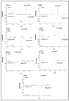
Urushiols are the allergenic components of Toxicodendron radicans (poison ivy) as well as other Toxicodendron species. They are alk-(en)-yl catechol derivatives with a 15- or 17-carbon side chain having different degrees of unsaturation. Although several methods have been developed for analysis of urushiols in plant tissues, the in situ localization of the different urushiol congeners has not been reported. Here, we report on the first analysis of urushiols in poison ivy stems by matrix-assisted laser desorption/ionization-mass spectrometry imaging (MALDI-MSI). Our results show that the urushiol congeners with 15-carbon side chains are mainly localized to the resin ducts, while those with 17-carbon side chains are widely distributed in cortex and vascular tissues. The presence of these urushiols in stem extracts of poison ivy seedlings was confirmed by GC-MS. These novel findings provide new insights into the spatial tissue distribution of urushiols that might be biosynthetically or functionally relevant.
Keywords: MALDI-MSI; Toxicodendron radicans; in situ localization; poison ivy; urushiols.
Conflict of interest statement
The authors declare no conflict of interest.
Figures


Similar articles
-
Accession-Level Differentiation of Urushiol Levels, and Identification of Cardanols in Nascent Emerged Poison Ivy Seedlings.Molecules. 2019 Nov 20;24(23):4213. doi: 10.3390/molecules24234213. Molecules. 2019. PMID: 31757036 Free PMC article.
-
Rapid detection of urushiol allergens of Toxicodendron genus using leaf spray mass spectrometry.Analyst. 2012 Mar 7;137(5):1082-4. doi: 10.1039/c2an16077c. Epub 2012 Jan 17. Analyst. 2012. PMID: 22249516
-
Urushiol Compounds Detected in Toxicodendron-Labeled Consumer Products Using Mass Spectrometry.Dermatitis. 2020 Mar/Apr;31(2):134-139. doi: 10.1097/DER.0000000000000544. Dermatitis. 2020. PMID: 32168145 Free PMC article.
-
Is it, or isn't it? Poison ivy look-a-likes.Am J Contact Dermat. 2000 Jun;11(2):104-10. doi: 10.1053/ac.2000.6337. Am J Contact Dermat. 2000. PMID: 10908180 Review.
-
Evaluation and Management of Toxicodendron Dermatitis in the Emergency Department: A Review of Current Practices.Wilderness Environ Med. 2023 Sep;34(3):388-392. doi: 10.1016/j.wem.2023.03.001. Epub 2023 Apr 27. Wilderness Environ Med. 2023. PMID: 37120383 Review.
Cited by
-
The chromosome-level genome for Toxicodendron vernicifluum provides crucial insights into Anacardiaceae evolution and urushiol biosynthesis.iScience. 2022 Jun 2;25(7):104512. doi: 10.1016/j.isci.2022.104512. eCollection 2022 Jul 15. iScience. 2022. PMID: 35733792 Free PMC article.
-
Poison ivy hairy root cultures enable a stable transformation system suitable for detailed investigation of urushiol metabolism.Plant Direct. 2020 Aug 7;4(8):e00243. doi: 10.1002/pld3.243. eCollection 2020 Aug. Plant Direct. 2020. PMID: 32783021 Free PMC article.
-
5-n-Alkylresorcinol Profiles in Different Cultivars of Einkorn, Emmer, Spelt, Common Wheat, and Tritordeum.J Agric Food Chem. 2021 Dec 1;69(47):14092-14102. doi: 10.1021/acs.jafc.1c05451. Epub 2021 Nov 18. J Agric Food Chem. 2021. PMID: 34793147 Free PMC article.
-
Sequencing and De Novo Assembly of the Toxicodendron radicans (Poison Ivy) Transcriptome.Genes (Basel). 2017 Nov 10;8(11):317. doi: 10.3390/genes8110317. Genes (Basel). 2017. PMID: 29125533 Free PMC article.
-
Accession-Level Differentiation of Urushiol Levels, and Identification of Cardanols in Nascent Emerged Poison Ivy Seedlings.Molecules. 2019 Nov 20;24(23):4213. doi: 10.3390/molecules24234213. Molecules. 2019. PMID: 31757036 Free PMC article.
References
-
- Epstein W. Poison oak and poison ivy dermatitis as an occupational problem. Cutis. 1974;13:544–548.
-
- Majima R., Chō S. Über einen hauptbestandteil des japanischen lackes. Eur. J. Inorg. Chem. 1907;40:4390–4393. doi: 10.1002/cber.19070400472. - DOI
-
- Majima R. Über den hauptbestandteil des japan-lacks, viii. Mitteilung: Stellung der doppelbindungen in der seitenkette des urushiols und beweisführung, daß das urushiol eine mischung ist. Eur. J. Inorg. Chem. 1922;55:172–191. doi: 10.1002/cber.19220550123. - DOI
-
- Symes W.F., Dawson C.R. Poison ivy “urushiol”. J. Am. Chem. Soc. 1954;76:2959–2963. doi: 10.1021/ja01640a030. - DOI
MeSH terms
Substances
LinkOut - more resources
Full Text Sources
Other Literature Sources
Research Materials
Miscellaneous

