Review
. 2017 Apr 29;22(5):713.
doi: 10.3390/molecules22050713.

Heparin, Heparan Sulphate and the TGF-β Cytokine Superfamily

Affiliations
Review

Heparin, Heparan Sulphate and the TGF-β Cytokine Superfamily

Chris C Rider et al. Molecules. .

Abstract

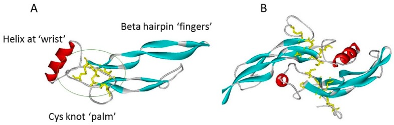
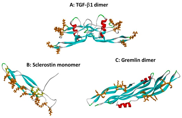
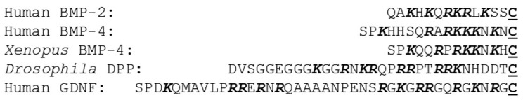
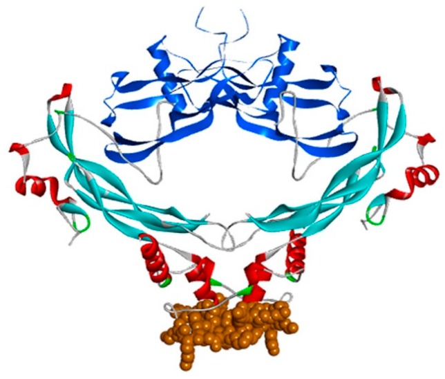
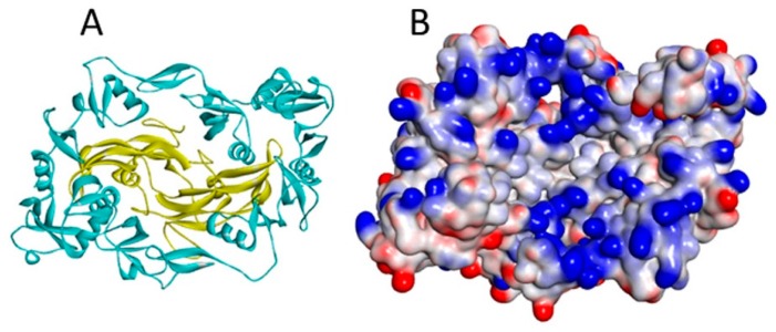
Of the circa 40 cytokines of the TGF-β superfamily, around a third are currently known to bind to heparin and heparan sulphate. This includes TGF-β1, TGF-β2, certain bone morphogenetic proteins (BMPs) and growth and differentiation factors (GDFs), as well as GDNF and two of its close homologues. Experimental studies of their heparin/HS binding sites reveal a diversity of locations around the shared cystine-knot protein fold. The activities of the TGF-β cytokines in controlling proliferation, differentiation and survival in a range of cell types are in part regulated by a number of specific, secreted BMP antagonist proteins. These vary in structure but seven belong to the CAN or DAN family, which shares the TGF-β type cystine-knot domain. Other antagonists are more distant members of the TGF-β superfamily. It is emerging that the majority, but not all, of the antagonists are also heparin binding proteins. Any future exploitation of the TGF-β cytokines in the therapy of chronic diseases will need to fully consider their interactions with glycosaminoglycans and the implications of this in terms of their bioavailability and biological activity.

Keywords: BMP antagonists; GDNF; TGF-β; bone morphogenetic protein (BMP); gremlin; growth and differentiation factor (GDF); heparan sulphate; heparin; noggin; sclerostin.


Conflict of interest statement

The authors declare no conflict of interest.

Figures

Figure 1
Figure 1
The TGF-β superfamily cystine knot fold, as typified by TGF-β1 (co-ordinates from 1KLC.pdb). Protein chains are shown in ribbon format: β-strands are blue, helices red, turns green; cystines are shown in yellow stick format. (A) TGF-β1 monomer showing the “hand” structures, with the cystine knot indicated by a green ellipse. (B) TGF-β1 dimer, the “wrist” of each hand is cupped in the other subunit. The view of the dimer is rotated by 90° with respect to the plane of 1A. The interchain disulphide bridge is visible in the centre of the structure.
Figure 2
Figure 2
Heparin binding sites on TGF-β superfamily cytokines. Protein chains are shown as in Figure 1, and heparin binding site basic residues are shown in brown stick format. (A) The dimer of TGF-β1 (co-ordinates from 1KLC.pdb). Residues K25, R26, K31, K37, R94 and R97 form a discontinuous heparin binding site at the tips of the “fingers” [13]. (B) Sclerostin monomer (one of the NMR ensemble in 2K8P.pdb) with heparin-binding residues in brown. Cystine residues are shown in yellow stick format. Loop 3 (the second β-strand loop) and Loop 2 are both involved; residues K99, R102, R114, R116, R119, R131, R133, K134, R136, K142, K144 and R145 form a linear heparin binding site capable of accommodating a heparin dodecamer [26]. (C) The dimer of the CAN BMP antagonist gremlin (co-ordinates from 5AEJ.pdb). Here, the heparin binding residues are located largely along the second “finger”, as for sclerostin; in the dimer both copies of the heparin binding site are on the concave face. Residues K145, K147, K148, K167, K168, K169, K174 and R177 have been identified as forming the heparin binding site [20]. The mode of dimerisation and the location of the heparin binding site both differ from those of TGF-β1.
Figure 3
Figure 3
N-terminal sequences of some heparin binding BMPs. The sequences are shown ending with the first cysteine (shown in bold underlined font at the right hand side) of the knot domain. The basic residues lysine and arginine are highlighted in bold italics, and sequence regions experimentally implicated in heparin binding are boxed.
Figure 4
Figure 4
The heparin binding site of noggin in the noggin-BMP-7 complex (co-ordinates from 1M4U.pdb). Noggin is shown as described in Figure 1, with amino acids 133–144, encompassing a cluster of eight basic arginine and lysine residues, shown in brown CPK format; BMP-7 is shown in blue ribbon format.
Figure 5
Figure 5
(A) The myostatin (yellow ribbon)/follistatin (turquoise ribbon) complex (3HH2.pdb); and (B) the same complex shown as a surface coloured according to interpolated charge (positive is blue, negative is red). Though myostatin does not have a heparin binding site, basic residues on its surface are located close to the follistatin binding site in the complex, increasing total affinity for heparin [40].

References

    1. Watabe T., Miyazono K. Roles of TGF-beta family signaling in stem cell renewal and differentiation. Cell Res. 2009;19:103–115. doi: 10.1038/cr.2008.323. - DOI - PubMed
    1. Fujio K., Komai T., Inoue M., Morita K., Okamura T., Yamamoto K. Revisiting the regulatory roles of the TGF-beta family of cytokines. Autoimmun. Rev. 2016;15:917–922. doi: 10.1016/j.autrev.2016.07.007. - DOI - PubMed
    1. Rodgarkia-Dara C., Vejda S., Erlach N., Losert A., Bursch W., Berger W., Schulte-Hermann R., Grusch M. The activin axis in liver biology and disease. Mutat. Res. 2006;613:123–137. doi: 10.1016/j.mrrev.2006.07.002. - DOI - PubMed
    1. Saarma M. GDNF—A stranger in the TGF-beta superfamily? Eur. J. Biochem. 2000;267:6968–6971. doi: 10.1046/j.1432-1327.2000.01826.x. - DOI - PubMed
    1. Rider C.C., Mulloy B. Bone morphogenetic protein and growth differentiation factor cytokine families and their protein antagonists. Biochem. J. 2010;429:1–12. doi: 10.1042/BJ20100305. - DOI - PubMed