Review
. 2017 May 1;18(5):956.
doi: 10.3390/ijms18050956.

Association of Extracellular Membrane Vesicles with Cutaneous Wound Healing

Affiliations
Review

Association of Extracellular Membrane Vesicles with Cutaneous Wound Healing

Uyen Thi Trang Than et al. Int J Mol Sci. .

Abstract

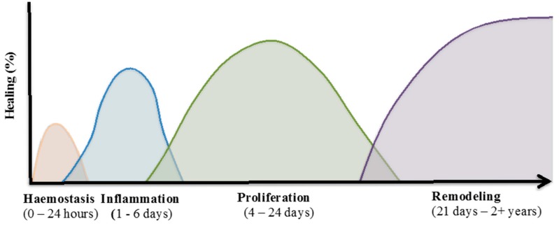
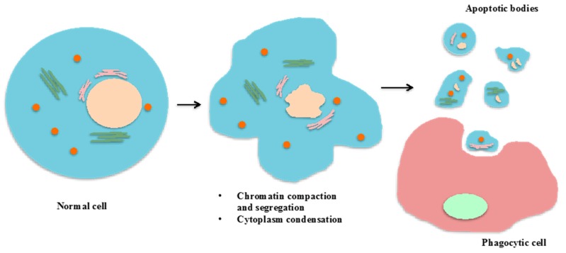
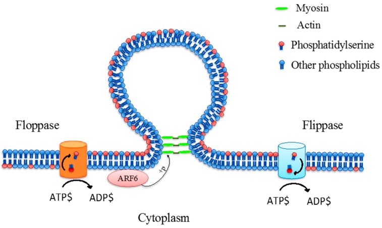
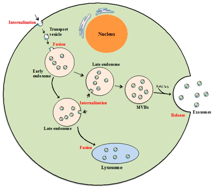
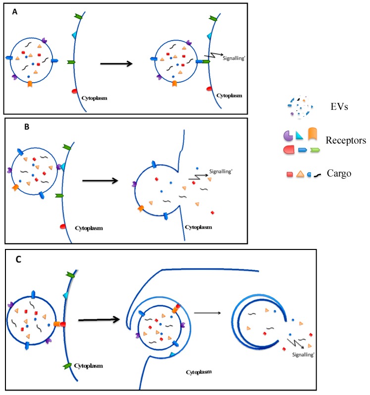
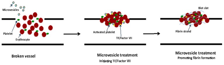
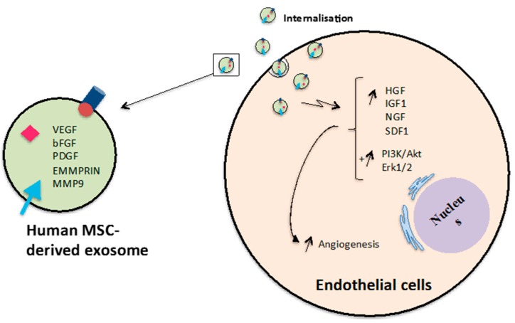
Extracellular vesicles (EVs) are membrane-enclosed vesicles that are released into the extracellular environment by various cell types, which can be classified as apoptotic bodies, microvesicles and exosomes. EVs have been shown to carry DNA, small RNAs, proteins and membrane lipids which are derived from the parental cells. Recently, several studies have demonstrated that EVs can regulate many biological processes, such as cancer progression, the immune response, cell proliferation, cell migration and blood vessel tube formation. This regulation is achieved through the release and transport of EVs and the transfer of their parental cell-derived molecular cargo to recipient cells. This thereby influences various physiological and sometimes pathological functions within the target cells. While intensive investigation of EVs has focused on pathological processes, the involvement of EVs in normal wound healing is less clear; however, recent preliminarily investigations have produced some initial insights. This review will provide an overview of EVs and discuss the current literature regarding the role of EVs in wound healing, especially, their influence on coagulation, cell proliferation, migration, angiogenesis, collagen production and extracellular matrix remodelling.

Keywords: angiogenesis; apoptotic bodies; endothelial cells; exosomes; extracellular membrane vesicles; keratinocytes; microvesicles; migration; proliferation; wound healing.


Conflict of interest statement

The authors declare no conflict of interest.

Figures

Figure 1
Figure 1
Wound healing process. The normal tissue repair process is comprised of continuous and overlapping phases. These four phases include: (i) Haemostasis; (ii) inflammation; (iii) proliferation; and (iv) remodelling. Each phase consists of different cellular events which requires the interplay of multiple cell populations [22].
Figure 2
Figure 2
Formation of apoptotic bodies and clearance by phagocytosis. Formation of apoptotic bodies includes the condensation and segregation of the nucleus, and the deterioration and blebbing of the plasma membrane. The result of these processes is a separation of the cellular contents into membrane-enclosed vesicles which can be cleared by phagocytic cells.
Figure 3
Figure 3
Phospholipid translocase activity via floppase and flippase which translocates phosphatidylserine and other phospholipids from the inner leaflet to the outer leaflet, and outer leaflet to inner leaflet, respectively, during microvesicle formation. These processes are adenosine triphosphate (ATP)-dependant [28,30,32,33].
Figure 4
Figure 4
Exosome biogenesis. Beginning from internalization of membrane proteins and lipid complexes by endocytosis, endocytotic vesicles are delivered to early endosomes, which fuse with each other resulting in formation of late endosomes/multivesicular bodies (MVB). MVBs either release exosomes by fusion with the cellular membrane, or their contents are degraded if they fuse with lysosomes [40,42,43]. The key steps of the exosomal formation and development process are highlighted in red.
Figure 5
Figure 5
Interaction of EVs with target cells: (A) intracellular signalling due to EV membrane ligand cell surface receptor interactions [53,54]; (B) direct membrane fusion which induces cell function through release of EV cargo into target cells [55,56,57,58]; and (C) internalisation of EVs into target cells, prior to the release of their cargo into the recipient cell cytoplasm inducing functional effects [21,59].
Figure 6
Figure 6
Promotion of coagulation by TF-barring microvesicle treatment. Rapid coagulation is triggered by the initiating TF/Factor VII and promotion of fibrin strand formation [98,99].
Figure 7
Figure 7
Promotion of angiogenesis by MSC-derived exosomes. Exosomes released from human MSCs can induce expression of genes and activate PI3K/Akt and Erk1/2 signalling pathways in endothelial cells leading to promotion tube formation and newly formed vessels.

Similar articles

Cited by

References

    1. Baietti M.F., Zhang Z., Mortier E., Melchior A., Degeest G., Geeraerts A., Ivarsson Y., Depoortere F., Coomans C., Vermeiren E. Syndecan-syntenin-ALIX regulates the biogenesis of exosomes. Nat. Cell Biol. 2012;14:677–685. doi: 10.1038/ncb2502. - DOI - PubMed
    1. Meister G., Landthaler M., Peters L., Chen P.Y., Urlaub H., Lührmann R., Tuschl T. Identification of novel argonaute-associated proteins. Curr. Biol. 2005;15:2149–2155. doi: 10.1016/j.cub.2005.10.048. - DOI - PubMed
    1. Valadi H., Ekström K., Bossios A., Sjöstrand M., Lee J.J., Lötvall J.O. Exosome-mediated transfer of mRNAs and microRNAs is a novel mechanism of genetic exchange between cells. Nat. Cell Biol. 2007;9:654–659. doi: 10.1038/ncb1596. - DOI - PubMed
    1. Vlassov A.V., Magdaleno S., Setterquist R., Conrad R. Exosomes: Current knowledge of their composition, biological functions, and diagnostic and therapeutic potentials. Biochim. Biophys. Acta. 2012;1820:940–948. doi: 10.1016/j.bbagen.2012.03.017. - DOI - PubMed
    1. Hao S., Bai O., Yuan J., Qureshi M., Xiang J. Dendritic cell-derived exosomes stimulate stronger CD8+ CTL responses and antitumor immunity than tumor cell-derived exosomes. Cell. Mol. Immunol. 2006;3:205–211. - PubMed