

The effect of dexmedetomidine on cerebral perfusion and oxygenation in healthy piglets with normal and lowered blood pressure anaesthetized with propofol-remifentanil total intravenous anaesthesia
- PMID: 28468670
- PMCID: PMC5415812
- DOI: 10.1186/s13028-017-0293-0
The effect of dexmedetomidine on cerebral perfusion and oxygenation in healthy piglets with normal and lowered blood pressure anaesthetized with propofol-remifentanil total intravenous anaesthesia
Abstract
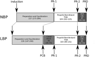
Background: During anaesthesia and surgery, in particular neurosurgery, preservation of cerebral perfusion and oxygenation (CPO) is essential for normal postoperative brain function. The isolated effects on CPO of either individual anaesthetic drugs or entire anaesthetic protocols are of importance in both clinical and research settings. Total intravenous anaesthesia (TIVA) with propofol and remifentanil is widely used in human neuroanaesthesia. In addition, dexmedetomidine is receiving increasing attention as an anaesthetic adjuvant in neurosurgical, intensive care, and paediatric patients. Despite the extensive use of pigs as animal models in neuroscience and the increasing use of both propofol-remifentanil and dexmedetomidine, very little is known about their combined effect on CPO in pigs with uninjured brains. This study investigates the effect of dexmedetomidine on CPO in piglets with normal and lowered blood pressure during background anaesthesia with propofol-remifentanil TIVA. Sixteen healthy female Danish pigs (crossbreeds of Danish Landrace, Yorkshire and Duroc, 25-34 kg) were used. Three animals were subsequently excluded. The animals were randomly allocated into one of two groups with either normal blood pressure (NBP, n = 6) or with induced low blood pressure (LBP, n = 7). Both groups were subjected to the same experimental protocol. Intravenous propofol induction was performed without premedication. Anaesthesia was maintained with propofol-remifentanil TIVA, and later supplemented with continuous infusion of dexmedetomidine. Assessments of cerebral perfusion obtained by laser speckle contrast imaging (LSCI) were related to cerebral oxygenation measures (PbrO2) obtained by an intracerebral Clark-type Licox probe.
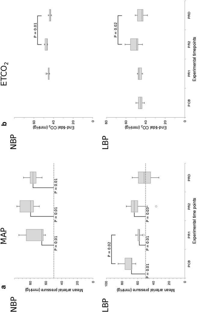
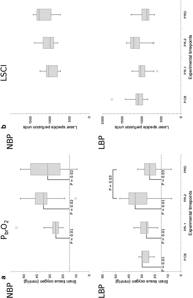
Results: Addition of dexmedetomidine resulted in a 32% reduction in median PbrO2 values for the LBP group (P = 0.03), but no significant changes in PbrO2 were observed for the NBP group. No significant changes in LSCI readings were observed in either group between any time points, despite a 28% decrease in the LBP group following dexmedetomidine administration. Caval block resulted in a significant (P = 0.02) reduction in median MAP from 68 mmHg (range 63-85) at PCB to 58 mmHg (range 53-63) in the LBP group, but no significant differences in either PbrO2 or LSCI were observed due to this intervention (P = 0.6 and P = 0.3 respectively).
Conclusions: Addition of dexmedetomidine to propofol-remifentanil TIVA resulted in a significant decrease in cerebral oxygenation (PbrO2) measurements in piglets with lowered blood pressure. Cerebral perfusion (LSCI) did not decrease significantly in this group. In piglets with normal blood pressure, no significant changes in cerebral perfusion or oxygenation were seen in response to addition of dexmedetomidine to the background anaesthesia.
Keywords: Cerebral oxygenation; Cerebral perfusion; Dexmedetomidine; Laser speckle contrast imaging; Licox; Propofol; Remifentanil; Swine.
Figures
Similar articles
-
The influence of norepinephrine and phenylephrine on cerebral perfusion and oxygenation during propofol-remifentanil and propofol-remifentanil-dexmedetomidine anaesthesia in piglets.Acta Vet Scand. 2018 Feb 8;60(1):8. doi: 10.1186/s13028-018-0362-z. Acta Vet Scand. 2018. PMID: 29422100 Free PMC article.
-
Effect of propofol and remifentanil on cerebral perfusion and oxygenation in pigs: a systematic review.Acta Vet Scand. 2016 Jun 22;58(1):42. doi: 10.1186/s13028-016-0223-6. Acta Vet Scand. 2016. PMID: 27334375 Free PMC article.
-
[Evaluation of heart rate variability for monitoring the depth of anaesthesia in dogs. Investigations based on total intravenous anaesthesia using propofol alone or in combination with dexmedetomidine or remifentanil].Tierarztl Prax Ausg K Kleintiere Heimtiere. 2015;43(1):1-10. doi: 10.15654/TPK-130744. Epub 2014 Nov 27. Tierarztl Prax Ausg K Kleintiere Heimtiere. 2015. PMID: 25428443 Clinical Trial. German.
-
Cerebral haemodynamic changes during propofol-remifentanil or sevoflurane anaesthesia: transcranial Doppler study under bispectral index monitoring.Br J Anaesth. 2006 Sep;97(3):333-9. doi: 10.1093/bja/ael169. Epub 2006 Jul 7. Br J Anaesth. 2006. PMID: 16829673 Clinical Trial.
-
[Total intravenous anesthesia. On the way to standard practice in pediatrics].Anaesthesist. 2003 Sep;52(9):763-77. doi: 10.1007/s00101-003-0560-5. Anaesthesist. 2003. PMID: 14504802 Review. German.
Cited by
-
Changes in brain connectivity and neurovascular dynamics during dexmedetomidine-induced loss of consciousness.bioRxiv [Preprint]. 2024 Nov 12:2024.10.04.616650. doi: 10.1101/2024.10.04.616650. bioRxiv. 2024. PMID: 39416182 Free PMC article. Preprint.
-
Changes in brain connectivity and neurovascular dynamics during dexmedetomidine-induced loss of consciousness.Commun Biol. 2025 Aug 20;8(1):1254. doi: 10.1038/s42003-025-08577-9. Commun Biol. 2025. PMID: 40835723 Free PMC article.
-
Effects of anesthesia on cerebral blood flow, metabolism, and neuroprotection.J Cereb Blood Flow Metab. 2018 Dec;38(12):2192-2208. doi: 10.1177/0271678X18789273. Epub 2018 Jul 16. J Cereb Blood Flow Metab. 2018. PMID: 30009645 Free PMC article. Review.
-
Analysis of Dexmedetomidine on the Quality of Awakening During Neurosurgery.Transl Neurosci. 2019 Jul 12;10:152-156. doi: 10.1515/tnsci-2019-0026. eCollection 2019. Transl Neurosci. 2019. PMID: 31410296 Free PMC article.
-
Application of Different Doses of Dexmedetomidine Combined with General Anesthesia in Anesthesia of Patients with Traumatic Tibiofibular Fractures and Its Effect on the Incidence of Adverse Reactions.J Healthc Eng. 2021 Dec 14;2021:3080098. doi: 10.1155/2021/3080098. eCollection 2021. J Healthc Eng. 2021. PMID: 34950440 Free PMC article.
References
-
- Venkatesh B. Monitoring cerebral perfusion and oxygenation: an elusive goal. Crit Care Resusc. 2005;7:195–199. - PubMed
-
- Dagal A, Lam AM. General considerations in neuroanaesthesia. In: Matta BF, Menon DK, Smith M, editors. Core topics in neuroanaesthesia and neurointensive care. edn. New York: Cambridge University Press; 2011. p.147–61.
-
- Engelhard K, Werner C. The effects of general anesthesia and variations in hemodynamics on cerebral perfusion. Appl Cardiopulm Pathophysiol. 2009;13:157–159.
-
- Ramani R, Todd M, Warner D. A680 The CBF response to isoflurane following brain injury in the rabbit. Anesthesiology. 1990;73:A680.
Publication types
MeSH terms
Substances
LinkOut - more resources
Full Text Sources
Other Literature Sources
Miscellaneous
