. 2017 May 3;10(1):219.
doi: 10.1186/s13071-017-2152-2.

CD3+CD4negCD8neg (double negative) T lymphocytes and NKT cells as the main cytotoxic-related-CD107a+ cells in lesions of cutaneous leishmaniasis caused by Leishmania (Viannia) braziliensis

Affiliations

CD3+CD4negCD8neg (double negative) T lymphocytes and NKT cells as the main cytotoxic-related-CD107a+ cells in lesions of cutaneous leishmaniasis caused by Leishmania (Viannia) braziliensis

Raquel Ferraz et al. Parasit Vectors. .

Abstract

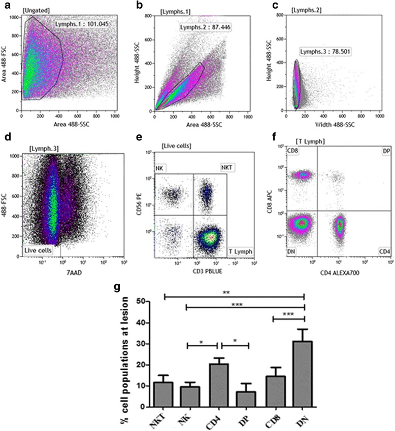
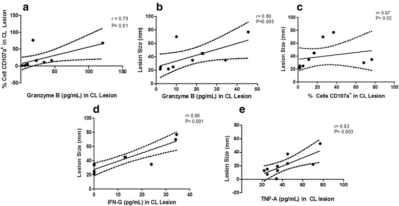
Background: Cutaneous leishmaniasis (CL) is caused by Leishmania (Viannia) braziliensis, which infects dermal macrophages and dendritic cells, causing an intense immune-mediated-tissue inflammation and a skin ulcer with elevated borders that can heal spontaneously or after antimonial therapy. The resolution of lesions depends on an adaptive immune response, and cytotoxic cells seem to have a fundamental role in this process. The aim of this study is to better understand the role of cytotoxicity mediated mechanisms that occur during the immune response in the CL lesion milieu, considering distinct cytotoxic-related CD107a+ cells, such as CD8+, CD4+, CD4neg CD8neg (double-negative, DN) and CD4+CD8+ (double-positive, DP) T lymphocytes, as well as NK and NKT cells.

Methods: Lesion derived cells were assessed for T cell subpopulations and NK cells, as well as CD107a expression by flow cytometry. In addition, cytometric bead array (CBA) was used to quantify cytokines and granzyme B concentrations in supernatants from macerated lesions.

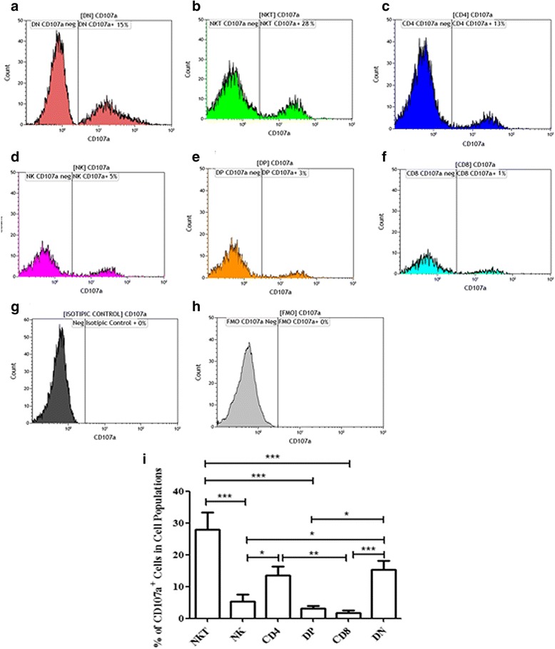
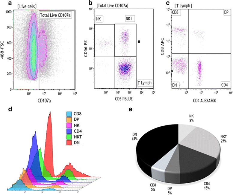
Results: Flow cytometry analyses revealed that NKT cells are the major CD107a-expressing cell population committed to cytotoxicity in CL lesion, although we also observed high frequencies of CD4+ and DN T cells expressing CD107a. Analysing the pool of CD107a+-cell populations, we found a higher distribution of DN T cells (44%), followed by approximately 25% of NKT cells. Interestingly, NK and CD8+ T cells represented only 3 and 4% of the total-CD107a+-cell pool, respectively.

Conclusions: The cytotoxicity activity that occurs in the lesion milieu of CL patients seems to be dominated by DN T and NKT cells. These findings suggest the need for a reevaluation of the role of classical-cytotoxic NK and CD8+ T cells in the pathogenesis of CL, implicating an important role for other T cell subpopulations.

Keywords: CD107a; Cytotoxicity; Double-negative T lymphocytes; Flow cytometry; Human cutaneous leishmaniasis; Leishmania (Viannia) braziliensis; Lesion; NKT cells.


Figures

Fig. 1
Fig. 1
Frequencies of NK and NKT cells; CD4+, CD8+, double positive (DP) and double negative (DN) CD3+ T-lymphocyte subpopulations. a-f Flow cytometry-representative protocol: Cells obtained from cutaneous leishmaniasis lesions were stained with anti-CD3, anti-CD4, anti-CD8, anti-CD56, anti-CD107a and 7-AAD. Lymphocytes region was created on forward (FSC) vs side (SSC) scatter dot-plot (a). Cells gated on [Lymphs.1] were analyzed through dot-plots SSC-Height vs SSC-Area (b) and SSC-Height vs SSC-Width (c) to exclude doublets and debris. d Dead cells (7-AAD+) were excluded from the analysis and a gate encompassing 7-AADneg cells was performed. e Based on this gate, NK cells (CD56+CD3neg), NKT (CD56+CD3+) and T lymphocytes (CD56negCD3+) were identified. f Based on T lymphocyte gate (T Lymph), CD4, CD8, double-negative (DN) and double-positive (DP) T lymphocytes were determined. g Bar graphs representing the mean ± SEM of percentages of NKT and NK cells; CD4+, DP, CD8+ and DN T lymphocytes obtained from cutaneous leishmaniasis lesions of 18 patients. Statistical analyses were performed using ANOVA test and Dunn’s post-hoc test. Results were considered significant with P < 0.05 (*P < 0.05; **P < 0.01; ***P < 0.001)
Fig. 2
Fig. 2
Frequencies of CD8+; CD4+; DP; DN lymphocytes; NK and NKT cells expressing CD107a from lesions of cutaneous leishmaniasis patients. Flow cytometry-representative analysis: a CD107a+DN T; b CD107a+NKT; c CD107a+CD4+ T; d CD107a+NK; e CD107a+DP T; f CD107a+CD8+ T cells. Isotype control (g) and fluorescence minus one (FMO) for CD107a staining (h). Electronic gates were created surrounding NKT, NK, CD4, DP, CD8 and DN cell populations (see Fig. 1). i The bars graph representing the mean ± SEM of percentages of NKT and NK cells; CD4+, DP, CD8+ and DN T lymphocytes expressing CD107a obtained from cutaneous leishmaniasis lesions of 18 patients. Statistical analyses between two groups were performed using Mann Whitney non-parametric t-test. Results were considered significant with P < 0.05 (*P < 0.05; **P < 0.01; ***P < 0.001)
Fig. 3
Fig. 3
Distribution of CD8+; CD4+; DP; DN lymphocytes; NK and NKT cells evaluated from the pool of CD107a+ cells. Flow cytometry-representative analysis to determine the distribution of cell populations in pool of CD107a+ cells. a FSC vs CD107a density-plot represents total percentage of CD107a+ cells based on [Live cells] gate (Fig. 1d). b CD3 vs CD56 density-plot gated on CD107a+ cells to define NK, NKT and T cells. c CD4 vs CD8 gated on CD3+ lymphocytes to define CD4, CD8, DP and DN T cells. d Overlay of CD8+; CD4+; DP; DN lymphocytes; NK and NKT cell histograms gated on CD107a+ cells. e Pie graph showing the mean data of NK and NKT cells; CD4+; DP, CD8+ and DN T lymphocytes (n = 18 CL lesions)
Fig. 4
Fig. 4
Correlation analysis of granzyme B, IFN-γ and TNF-α production, frequency of CD107+ cells and lesion size from lesions of CL patients (n = 10). Granzyme B, IFN-γ and TNF-α levels were quantified in culture media from macerated lesions biopsied from ten patients. All samples were prepared following CBA multiplex kit manual and the cytokines were detected within a range of 10–2500 pg/ml. We observed positive correlations between frequency of CD107a+ cells and granzyme B production (a); lesion size and granzyme B production (b); lesion size and frequency of CD107a+ cells in CL lesions (c); lesion size and IFN-γ production (d); and lesion size and TNF-α production (e). The central line represents median values and the graph show best fitted lines with 95% confidence interval. Statistical analyses were performed using Spearman’s test (r: correlation coefficient). Results were considered significant with P < 0.05. Each point represents one CL patient

Similar articles

Cited by

References

    1. WHO | Leishmaniasis. WHO. [cited 2015 Mar 25]. Available from: http://www.who.int/mediacentre/factsheets/fs375/en/
    1. De Oliveira-Neto MP, Mattos MS, Perez MA, Da-Cruz AM, Fernandes O, Moreira J, et al. American tegumentary leishmaniasis (ATL) in Rio de Janeiro State, Brazil: main clinical and epidemiologic characteristics. Int J Dermatol. 2000;39:506–14. doi: 10.1046/j.1365-4362.2000.00969.x. - DOI - PubMed
    1. Brelaz-de-Castro MCA, de Almeida AF, de Oliveira AP, de Assis-Souza M, da Rocha LF, Pereira VRA. Cellular immune response evaluation of cutaneous leishmaniasis patient cells stimulated with Leishmania (Viannia) braziliensis antigenic fractions before and after clinical cure. Cell Immunol. 2012;279:180–6. doi: 10.1016/j.cellimm.2012.11.006. - DOI - PubMed
    1. Bottrel RL, Dutra WO, Martins FA, Gontijo B, Carvalho E, Barral-Netto M, et al. Flow cytometric determination of cellular sources and frequencies of key cytokine-producing lymphocytes directed against recombinant LACK and soluble Leishmania antigen in human cutaneous leishmaniasis. Infect Immun. 2001;69:3232–9. doi: 10.1128/IAI.69.5.3232-3239.2001. - DOI - PMC - PubMed
    1. Bacellar O, Lessa H, Schriefer A, Machado P. Ribeiro de Jesus A, Dutra WO, et al. Up-regulation of Th1-type responses in mucosal leishmaniasis patients. Infect Immun. 2002;70:6734–40. doi: 10.1128/IAI.70.12.6734-6740.2002. - DOI - PMC - PubMed

MeSH terms

LinkOut - more resources