Review
. 2017 Jul 1;97(3):889-938.
doi: 10.1152/physrev.00038.2016.

LITTLE FISH, BIG DATA: ZEBRAFISH AS A MODEL FOR CARDIOVASCULAR AND METABOLIC DISEASE

Affiliations
Review

LITTLE FISH, BIG DATA: ZEBRAFISH AS A MODEL FOR CARDIOVASCULAR AND METABOLIC DISEASE

Philipp Gut et al. Physiol Rev. .

Abstract

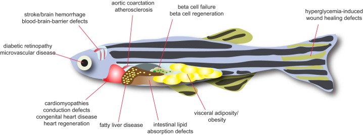
The burden of cardiovascular and metabolic diseases worldwide is staggering. The emergence of systems approaches in biology promises new therapies, faster and cheaper diagnostics, and personalized medicine. However, a profound understanding of pathogenic mechanisms at the cellular and molecular levels remains a fundamental requirement for discovery and therapeutics. Animal models of human disease are cornerstones of drug discovery as they allow identification of novel pharmacological targets by linking gene function with pathogenesis. The zebrafish model has been used for decades to study development and pathophysiology. More than ever, the specific strengths of the zebrafish model make it a prime partner in an age of discovery transformed by big-data approaches to genomics and disease. Zebrafish share a largely conserved physiology and anatomy with mammals. They allow a wide range of genetic manipulations, including the latest genome engineering approaches. They can be bred and studied with remarkable speed, enabling a range of large-scale phenotypic screens. Finally, zebrafish demonstrate an impressive regenerative capacity scientists hope to unlock in humans. Here, we provide a comprehensive guide on applications of zebrafish to investigate cardiovascular and metabolic diseases. We delineate advantages and limitations of zebrafish models of human disease and summarize their most significant contributions to understanding disease progression to date.


Figures

FIGURE 1.
FIGURE 1.
Size and shape of zebrafish research. A: percentage of publications involving the four most frequently used “lower” organisms Drosophila, C. elegans, Danio rerio, and Xenopus as a proportion of all articles published in MEDLINE. After the publication of the initial large-scale forward genetic screens in 1996, the zebrafish became increasingly popular as a model organism. B: percentage of NIH R01s involving zebrafish research is on the rise. [Graph modified from Lauer (199). Reprinted by permission from Macmillan Publishers Ltd. Data from Nature/The week in science: 5–11, August 2016; reproduced with permission from the NIH Office of Extramural Research/Open Mike blog.] C: geotagging authors’ affiliations from zebrafish papers in a MEDLINE search shows where in the world zebrafish research is taking place.
FIGURE 2.
FIGURE 2.
Advantages of zebrafish for biomedical research. A: zebrafish occupy a unique position in biomedical research as a vertebrate organism accessible to large-scale genetic and chemical screening. B: zebrafish are amenable to a broad spectrum of genomic, physiological, imaging, and small-molecule screening approaches.
FIGURE 3.
FIGURE 3.
Genome manipulations. A: overview of technologies to manipulate the zebrafish genome. B: guidelines for the correct use of morpholino (MO)-mediated gene knockdown technology. The current recommendation to identify a MO with minimal off-target effects is to test it in a null mutant background, as it should not induce any additional phenotypes.
FIGURE 4.
FIGURE 4.
Overview of decision roadmap to model genetic disease in zebrafish. Complex and monogenic human diseases can be modeled in zebrafish using different strategies, including genome editing, morpholinos, or transgenic technologies. Several considerations have to be made such as the conservation of genes between zebrafish and human, the timeline of generating genetically modified zebrafish, existence of more suitable animal models, and the availability of meaningful assays to match phenotypic findings to clinical symptoms.
FIGURE 5.
FIGURE 5.
Cardiovascular and metabolic diseases studied in zebrafish. Zebrafish have a similar anatomy as that of higher vertebrates. Tissue-intrinsic pathological processes can be studied for a broad range of human cardiovascular and metabolic diseases.
FIGURE 6.
FIGURE 6.
Development of cardiovascular physiology. Cardiac peristalsis and blood circulation can be detected by 24 hpf, making the zebrafish a prime model to study early events of cardiovascular development and physiology. The full complement of anatomical and physiological characteristics of an adult heart develops gradually and is considered to be complete by around 6 wk post fertilization. Analogous to cardiac development, the different cell types of the vascular system develop progressively; for example, a fully mature blood-brain barrier is not established before 14 dpf.
FIGURE 7.
FIGURE 7.
Modeling and phenotyping of cardiac disease. A: schematic of adult zebrafish EKG measurement. An anesthetized zebrafish is positioned ventral side up on a damp sponge; a gill perfusion pump may be used. Needle electrodes are positioned under the skin, and the EKG signal is amplified, filtered, and acquired using standard equipment (246). B: the zebrafish model for Long QT Syndrome Type 2 (LQT2) has a clinically relevant phenotype. Human LQT2 mutations are autosomal dominant and cause a long QT interval on EKG. Analogously, heterozygous kcnh6 adults also demonstrate prolonged QT interval compared with wild-type siblings. C: homozygous kcnh6−/− embryos are amenable to detailed phenotyping, showing absence of ventricular conduction by optical mapping. Wild-type and mutant embryos were crossed into the Tg(myl7:gCaMP) fluorescent calcium sensor transgenic background, and imaged using selective plane illumination microscopy. Hearts were stopped to facilitate imaging of conduction. Fluorescence intensity over time was plotted from several regions of interest from the embryos’ atrium and ventricle, showing that cyclic fluorescence, and therefore conduction, was absent in the mutant ventricle. This observation was confirmed by patch-clamping the embryonic hearts. [B and C from Arnaout et al. (9).]
FIGURE 8.
FIGURE 8.
Development of metabolic regulation in zebrafish. A: timeline of metabolic characteristics throughout development. Glucose levels are highly dynamic during development and are regulated at early stages by endogenous glucose production, glucose utilization, as well as hormones, as in mammalian glucoregulation. Lipid homeostasis is determined at early stages by release of stored lipids within the yolk sac. The first adipose cells appear after feeding commences, and larger fat depots are not formed before ~30 dpf. B: energy substrates in the yolk sustain growth and metabolism, but are depleted around 4.5 dpf. If larvae are not fed by this time, they switch to a fasted state, and metabolic adaptations set in that are reminiscent of mammalian energy-sensing mechanisms. This defined yolk-feeding to fasting transition can be leveraged to monitor energy-sensing mechanisms. In addition, feeding of zebrafish larvae can be used to monitor adipocyte formation or intestinal lipid processing [Modified from Schlegel and Gut (341).]
FIGURE 9.
FIGURE 9.
Zebrafish assist in all drug discovery and development phases. Fundamental and preclinical research using zebrafish generate new targets and help to validate predicted targets in vivo. Cornerstones of the traditional R&D pipeline including lead optimization, toxicology testing, and long-term efficacy as well as safety studies are being developed in preclinical zebrafish assays. Drug repurposing can match existing drugs with potentially new clinical indications through screening in disease models. Incorporating whole-organism testing into traditional R&D pipelines early in the process can help critical decision-making and avoid late-stage failure of new chemical entities.
FIGURE 10.
FIGURE 10.
From human disease to therapies using zebrafish. In many cases, disease models can be used to develop screening paradigms for suppressor molecules of pathological hallmarks. This emerging concept has been shown to facilitate the transition from preclinical to clinical trials, in particular in areas of unmet clinical need through screening of clinically approved drug collections. Advantages and disadvantages of available chemical libraries are summarized in the box.

Similar articles

Cited by

References

    1. Alvarez Y, Chen K, Reynolds AL, Waghorne N, O’Connor JJ, Kennedy BN. Predominant cone photoreceptor dysfunction in a hyperglycaemic model of non-proliferative diabetic retinopathy. Dis Model Mech 3: 236–245, 2010. doi:10.1242/dmm.003772. - DOI - PubMed
    1. Amsterdam A, Burgess S, Golling G, Chen W, Sun Z, Townsend K, Farrington S, Haldi M, Hopkins N.. A large-scale insertional mutagenesis screen in zebrafish. Genes Dev 13: 2713–2724, 1999. doi:10.1101/gad.13.20.2713. - DOI - PMC - PubMed
    1. Anderson JL, Carten JD, Farber SA. Zebrafish lipid metabolism: from mediating early patterning to the metabolism of dietary fat and cholesterol. Methods Cell Biol 101: 111–141, 2011. doi:10.1016/B978-0-12-387036-0.00005-0. - DOI - PMC - PubMed
    1. Anderson KV, Jürgens G, Nüsslein-Volhard C. Establishment of dorsal-ventral polarity in the Drosophila embryo: genetic studies on the role of the Toll gene product. Cell 42: 779–789, 1985. doi:10.1016/0092-8674(85)90274-0. - DOI - PubMed
    1. Andersson O, Adams BA, Yoo D, Ellis GC, Gut P, Anderson RM, German MS, Stainier DY. Adenosine signaling promotes regeneration of pancreatic β cells in vivo. Cell Metab 15: 885–894, 2012. doi:10.1016/j.cmet.2012.04.018. - DOI - PMC - PubMed

Publication types

MeSH terms

Substances