. 2017 Jun 5;216(6):1715-1730.
doi: 10.1083/jcb.201612123. Epub 2017 May 3.

A voltage-dependent K+ channel in the lysosome is required for refilling lysosomal Ca2+ stores

Affiliations

A voltage-dependent K+ channel in the lysosome is required for refilling lysosomal Ca2+ stores

Wuyang Wang et al. J Cell Biol. .

Abstract

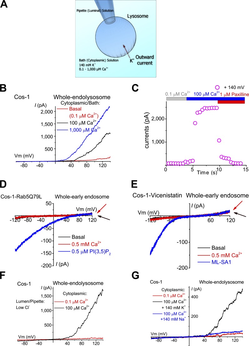
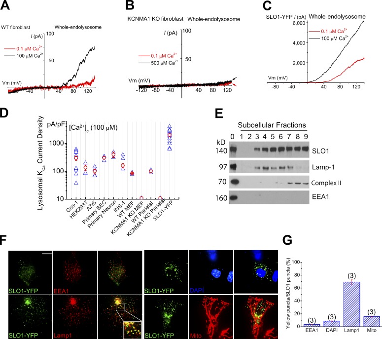
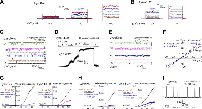
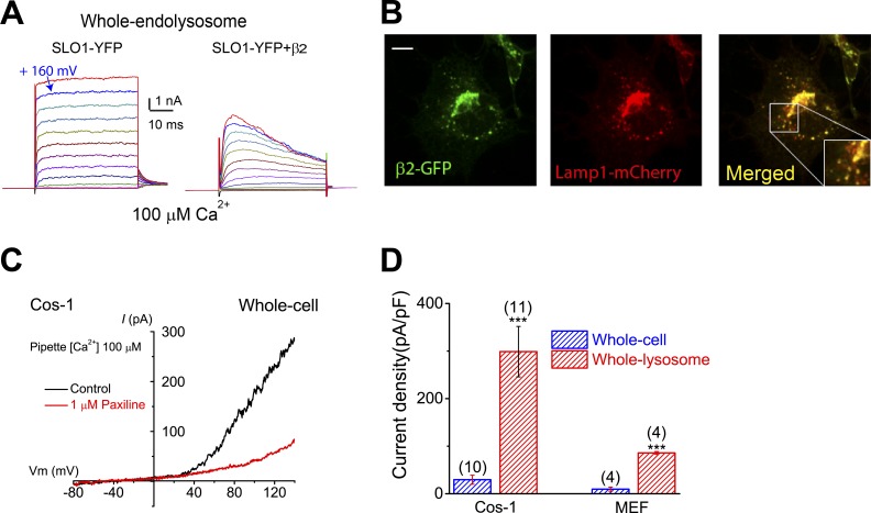
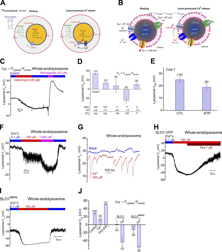
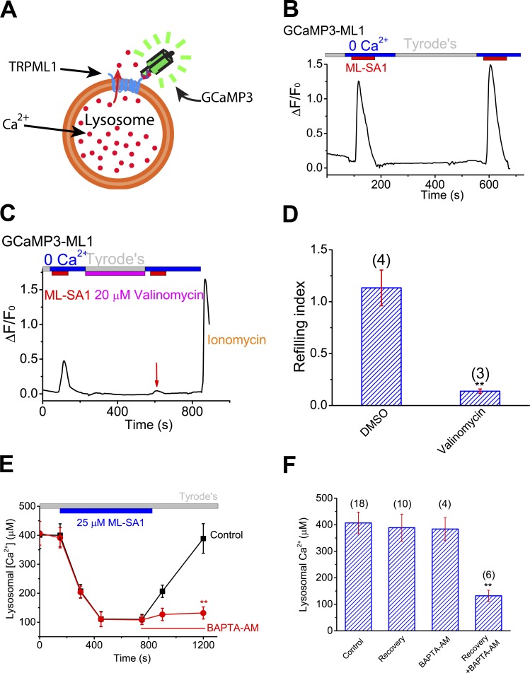
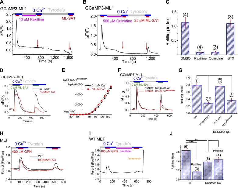
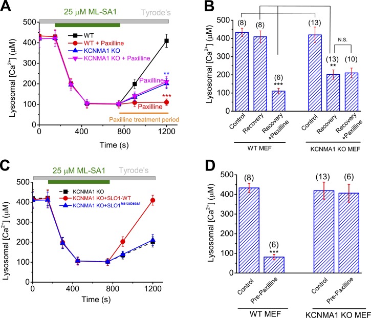
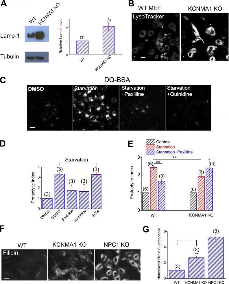
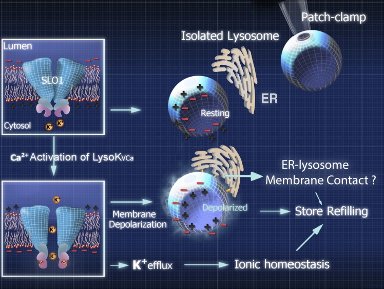
The resting membrane potential (Δψ) of the cell is negative on the cytosolic side and determined primarily by the plasma membrane's selective permeability to K+ We show that lysosomal Δψ is set by lysosomal membrane permeabilities to Na+ and H+, but not K+, and is positive on the cytosolic side. An increase in juxta-lysosomal Ca2+ rapidly reversed lysosomal Δψ by activating a large voltage-dependent and K+-selective conductance (LysoKVCa). LysoKVCa is encoded molecularly by SLO1 proteins known for forming plasma membrane BK channels. Opening of single LysoKVCa channels is sufficient to cause the rapid, striking changes in lysosomal Δψ. Lysosomal Ca2+ stores may be refilled from endoplasmic reticulum (ER) Ca2+ via ER-lysosome membrane contact sites. We propose that LysoKVCa serves as the perilysosomal Ca2+ effector to prime lysosomes for the refilling process. Consistently, genetic ablation or pharmacological inhibition of LysoKVCa, or abolition of its Ca2+ sensitivity, blocks refilling and maintenance of lysosomal Ca2+ stores, resulting in lysosomal cholesterol accumulation and a lysosome storage phenotype.


Figures

Figure 1.
Figure 1.
Ca2+-activated K+ currents were detected in lysosomes, but not in early endosomes. (A) Whole-endolysosome patch-clamp configuration. The pipette (extracellular/luminal) solution was a standard Na+-based external solution (Tyrode’s) adjusted to pH 4.6 to mimic the acidic environment of the lysosome lumen. The bath (internal/cytoplasmic) solution was a K+-based solution (140 mM K+) with free [Ca2+] ranging from 0.1 to 1,000 µM as indicated in each experiment. The arrow indicates the direction of K+ ion flow into the endolysosome (defined as the outward current). (B) Bath application of 100 or 1,000 µM Ca2+ activated outwardly rectifying currents in an enlarged vacuole from a Cos-1 cell pretreated with vacuolin-1. The currents were elicited by repeated voltage ramps (−100 to 140 mV; 400 ms; only partial voltage ranges are shown) with a 4-s interramp interval. (C) Time courses of current activation by Ca2+ in an enlarged vacuole isolated from a Cos-1 cell. (D and E) In isolated early endosomes that were enlarged by Rab5-Q79L overexpression (D) or vicenistatin (E), Ca2+ (500 µM) failed to activate measurable whole-endosome outward currents. PI(3,5)P2 (0.5 µM) or ML-SA1 (20 µM) readily activated TPC-like (D) or ML1-like (E) currents, respectively. (F) Representative traces of Ca2+-activated outward currents with a pipette/luminal solution containing low [Cl] (11 mM versus 156 mM in G. (G) Substitution of cytoplasmic K+ with Na+ abolished Ca2+-activated outward currents.
Figure 2.
Figure 2.
SLO1 mediates Ca2+-activated K+ currents in the lysosomes of excitable and nonexcitable cells. (A) Whole-endolysosome Ca2+-activated outward currents in a WT MEF. (B) Whole-endolysosome Ca2+-activated outward currents in a KCNMA1 KO (Kcnma1−/− or Slo1−/−) MEF. Note the lack of background outward currents typically seen in whole-cell recordings. (C) Whole-endolysosome Ca2+-activated currents in a SLO1-YFP–expressing Cos-1 cell (Lyso-SLO1). (D) Summary of whole-endolysosome Ca2+- and voltage-activated K+ currents (LysoKVCa) in a variety of cell types, including Cos-1 cells, HEK293T cells, A7r5 smooth muscle cell lines, mouse BECs, cultured mouse cortical neurons, INS-1 pancreatic cell lines, WT and KCNMA1 KO MEFs, WT and KCNMA1 KO parietal cells, and SLO1-YFP-expressing Cos-1 cells. Both individual (blue) and mean (red; ± SEM) current densities are shown for each cell type (n = 3–15 patches). (E) Subcellular fractionation analysis revealed enrichment of SLO1 proteins in organellar fractions containing Lamp-1 or Complex-II (a mitochondrial marker). Subcellular fractionations (1–9) were obtained by gradient-based ultracentrifugation. Cell lysates were included as controls (fraction 0). (F and G) Colocalization analyses of SLO1-YFP with Lamp1, MitoTracker, EEA1 (an early endosomal marker), and DAPI (a nuclear marker). Bar, 10 µm. Error bars indicate SEM.
Figure 3.
Figure 3.
LysoKVCa is a lysosomal large-conductance K+ channel that is dually activated by Ca2+ and voltage. (A) LysoKVCa currents were activated by cytoplasmic Ca2+ ([Ca2+]C; 0.1, 100, and 1,000 µM). After a 20-ms prepulse at −60 mV, currents were elicited by voltage steps from −80 to 180 mV in 20-mV increments. (B) Activation of Lyso-SLO1 by Ca2+ and voltage steps. (C) [Ca2+]C-dependent single-channel LysoKVCa currents from a cytoplasmic-side-out patch. The open and closed states are indicated by O and C, respectively. (D) [Ca2+]C dependence of macroscopic Lyso-SLO1 currents. (E) Voltage-dependent openings of single LysoKVCa channels at [Ca2+]C of 100 µM in a cytoplasmic-side-out patch with symmetric (luminal/cytoplasmic) K+ (140 mM) solutions. (F) Unitary single-channel chord conductances of LysoKVCa and Lyso-SLO1. (G) LysoKVCa and Lyso-SLO1 currents were blocked by the BK-specific inhibitor paxilline (1 µM). (H) Quinidine (500 µM), a SLO1/SLO3 channel inhibitor, blocked LysoKVCa and Lyso-SLO1 currents. (I) Single LysoKVCa currents were inhibited by bath application of 100 nM IBTX to a luminal-side-out patch.
Figure 4.
Figure 4.
LysoKVCa is preferentially expressed in the lysosome. (A) Step currents of Lyso-SLO1 (left) and Lyso-SLO1+ β2 (right) in the presence of 100 µM Ca2+. (B) Localization of β2-GFP in the Lamp1-positive compartments. Bar, 10 µm. (C) Whole-cell K+ currents sensitive to paxilline (1 µM) in nontransfected Cos-1 cells. The pipette solution contained 100 µM Ca2+. (D) Densities (mean ± SEM) of plasma membrane BK-like currents vs. LysoKVCa in Cos-1 cells and MEFs. Statistical comparisons were made with variance analysis (Student’s t test). ***, P < 0.001.
Figure 5.
Figure 5.
Regulation of lysosomal membrane potential by lysosomal K+ and LysoKVCa. (A) When the voltage or potential (ψ) of the extracellular solution is set to 0 mV by conventional definition, the ψ of the cytosol (ψCytosol) is approximately −70 mV. Hence the resting membrane potential (Δψ) of the cell is approximately −70 mV (cytoplasmic-side negative). At resting conditions, ψLumen is −100 mV and lysosomal Δψ (ψCytosol − ψLumen) is 30 mV. Upon activation of LysoKVCa, depending on the extent of LysoKVCa activation, lysosomal Δψ is reversed to −30 to −60 mV. (B) When ψCytosol is set to 0 mV, lysosomal Δψ at rest is 30 mV. Assuming >10-fold concentration gradients across lysosomal membranes for Na+ and K+ ([Na+]L >> [Na+]C and [K+]L << [K+]C), ENa is >57 mV and EK is <−57 mV. Likewise, EH is >149 mV. The resting Δψ is determined primarily by Na+ and H+ permeabilities. Upon Ca2+ activation of LysoKVCa, lysosomal Δψ is changed to −30 to −60 mV. (C) Effects of K+ (valinomycin) and H+ (niclosamide) ionophores on lysosomal Δψ in a Cos-1 cell in a current-clamp vacuole. (D) Summary of lysosomal Δψ under different ionic compositions in the luminal and cytoplasmic sides. Lysosomal Δψ was 20 – 30 mV under control conditions (lumen, 145 mM Na+, pH 4.6; cytosol, 140 mM K+, pH 7.4). Replacement of luminal Na+ with NMDG+ or increasing luminal pH from 4.6 to 7.4 led to a reduction in Δψ. (E) Resting lysosomal Δψ with or without ATP in the bath/cytoplasmic solutions. (F) Current-clamp recordings of lysosomal Δψ in a Cos-1 cell. (G) [Ca2+]C (100 µM) induced voltage transients in a current-clamped vacuole. Single-channel LysoKVCa currents from the same vacuole under the voltage-clamp configuration are shown in Fig. S5 H. (H and I) Lysosomal Δψ in enlarged vacuoles from SLO1-YFP-expressing (H) and SLO1R207Q-expressing (I) cells. LysoKVCa currents from the same patches under the voltage-clamp configuration are shown in Fig. S5, K and L. (J) Summary of lysosomal Δψ in various conditions. In D, E, and J, means ± SEM are shown.
Figure 6.
Figure 6.
Lysosomal K+ homeostasis and cytosolic Ca2+ increase are both required for lysosomal Ca2+ store refilling. (A) Lysosome-targeted genetically encoded Ca2+ indicator GCaMP3-ML1. (B) In HEK293 cells stably expressing GCaMP3-ML1 (HEK-GCaMP3-ML1 cells), lysosomal Ca2+ release, as indicated by elevated GCaMP3 fluorescence (F470), was induced by bath application of ML-SA1 (25 µM), a membrane-permeable ML1-specific agonist, in a 0 Ca2+ (<10 nM) external solution. Averaged responses of 15–30 cells in one coverslip are shown. (C) Short-term (5-min) application of valinomycin (20 µM) in the refilling phase abolished the second ML-SA1 response. (D) Effect of valinomycin on lysosome Ca2+ refilling. (E and F) Effect of BAPTA-AM treatment on recovery of lysosomal Ca2+ content upon ML-SA1 treatment in ML1-expressing HEK293 cells that were loaded with OG-BAPTA dextran dyes. Statistical comparisons were made with variance analysis (Student’s t test). **, P < 0.01. Error bars indicate SEM.
Figure 7.
Figure 7.
LysoKVCa and its Ca2+ sensitivity regulate the refilling of lysosomal Ca2+ stores. (A and B) Acute application of paxilline (A) and quinidine (B) abolished the second ML-SA1-induced responses. Prolonged washout for 10–15 min led to a partial recovery of the responses. (C) Lysosome Ca2+ refilling in HEK-GCaMP3-ML1 cells treated with paxilline, quinidine, and IBTX. (D) Compared with WT MEFs, ML-SA1–stimulated refilled responses were reduced in GCaMP7-ML1-expressing KCNMA1 KO MEFs. (E) Lyso-SLO1M513I/D898A currents at different concentrations of Ca2+ (0.1 and 10 µM). (F and G) Lysosomal refilling in GCaMP7-ML1–expressing KCNMA1 KO MEFs that were transfected WT SLO-mCherry and SLO1M513I/D898A-mCherry. (H) GPN-induced refilled (the second) Ca2+ response, measured with Fura-2 imaging, was reduced in KCNMA1 KO MEFs. (I) Paxilline effects in lysosomal refilling in WT MEF cells. (J) Mean refilling responses in WT and KCNMA1 KO MEF cells. Statistical comparisons were made with variance analysis (Student’s t test). **, P < 0.01; ***, P < 0.001. Error bars indicate SEM.
Figure 8.
Figure 8.
LysoKVCa is required for the maintenance of lysosomal Ca2+ contents. (A and B) Effects of paxilline on ML-SA1–induced changes in lysosomal Ca2+ contents in WT and KCNMA1 KO MEFs. (C) Effects of overexpressing WT SLO-mCherry and SLO1M513I/D898A-mCherry on lysosomal Ca2+ contents in HEK293 cells. (D) Effects of pretreatment with paxilline on lysosomal Ca2+contents in WT and KCNMA1 KO MEFs. Statistical comparisons were made with variance analysis (Student’s t test). **, P < 0.01; ***, P < 0.001; N.S., not significant. Error bars indicate SEM.
Figure 9.
Figure 9.
LysoKVCa is required for lysosome function. (A) Western blot analysis of Lamp1 expression in WT and KCNMA1 KO MEFs. (B) LysoTracker staining in WT and KCNMA1 KO MEFs. Bar, 10 µm. (C) Confocal imaging of DQ-red-BSA in starved Cos-1 cells (amino acid + serum starvation) in the presence of paxilline (10 µM), quinidine (500 µM), or IBTX (100 nM). Bar, 10 µm. (D) Normalized proteolytic index for starved Cos-1 cells treated with paxilline or quinidine. (E) Normalized proteolytic index for starved WT and KCNMA1 KO MEFs treated with paxilline. (F) Cholesterol levels, detected with filipin staining, in WT, KCNMA1 KO, and NPC1 KO MEFs. Bar, 50 µm. (G) Normalized filipin density in WT, KCNMA1 KO, and NPC1 KO MEFs. Means ± SEM are shown in A, D, E, and G. Statistical comparisons were made using variance analysis (ANOVA for D, E, and G). *, P < 0.05; **, P < 0.01.
Figure 10.
Figure 10.
A working model of the potential roles of LysoKVCa-mediated K+ flux in lysosomal dynamics and Ca2+ store refilling. Endolysosomes have a positive Δψ (Fig. 5, A and B) at rest. Upon juxta-lysosomal Ca2+ increase, LysoKVCa-mediated K+ flux causes reduction and reversal of Δψ toward EK (−57 mV, assuming a 10-fold concentration gradient). Changes in lysosomal Δψ may initiate the refilling of lysosomal Ca2+ stores via yet-to-be-defined mechanisms, such as formation of ER–lysosome membrane contact sites.

References

    1. Bandyopadhyay D., Cyphersmith A., Zapata J.A., Kim Y.J., and Payne C.K.. 2014. Lysosome transport as a function of lysosome diameter. PLoS One. 9:e86847 10.1371/journal.pone.0086847 - DOI - PMC - PubMed
    1. Bao L., Kaldany C., Holmstrand E.C., and Cox D.H.. 2004. Mapping the BKCa channel’s ‘Ca2+ bowl’: Side-chains essential for Ca2+ sensing. J. Gen. Physiol. 123:475–489. 10.1085/jgp.200409052 - DOI - PMC - PubMed
    1. Berg T.O., Strømhaug E., Løvdal T., Seglen O., and Berg T.. 1994. Use of glycyl-L-phenylalanine 2-naphthylamide, a lysosome-disrupting cathepsin C substrate, to distinguish between lysosomes and prelysosomal endocytic vacuoles. Biochem. J. 300:229–236. 10.1042/bj3000229 - DOI - PMC - PubMed
    1. Braun M., Ramracheya R., Bengtsson M., Zhang Q., Karanauskaite J., Partridge C., Johnson P.R., and Rorsman P.. 2008. Voltage-gated ion channels in human pancreatic beta-cells: Electrophysiological characterization and role in insulin secretion. Diabetes. 57:1618–1628. 10.2337/db07-0991 - DOI - PubMed
    1. Bright N.A., Davis L.J., and Luzio J.P.. 2016. Endolysosomes are the principal intracellular sites of acid hydrolase activity. Curr. Biol. 26:2233–2245. 10.1016/j.cub.2016.06.046 - DOI - PMC - PubMed

MeSH terms

Substances