. 2017 Jun 26;91(14):e00084-17.
doi: 10.1128/JVI.00084-17. Print 2017 Jul 15.

Regulation of Tacaribe Mammarenavirus Translation: Positive 5' and Negative 3' Elements and Role of Key Cellular Factors

Affiliations

Regulation of Tacaribe Mammarenavirus Translation: Positive 5' and Negative 3' Elements and Role of Key Cellular Factors

Sabrina Foscaldi et al. J Virol. .

Abstract

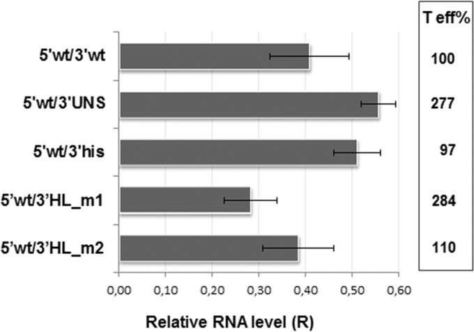
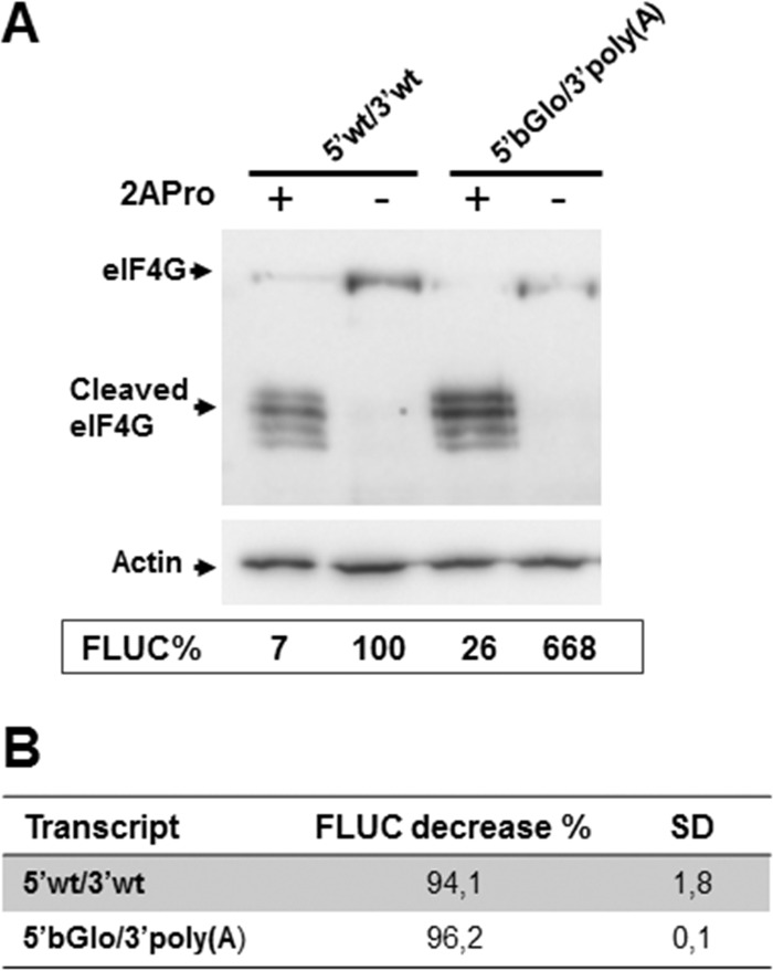
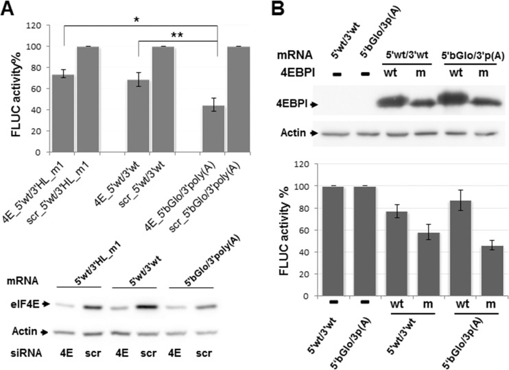
Mammarenaviruses are enveloped viruses with a bisegmented negative-stranded RNA genome that encodes the nucleocapsid protein (NP), the envelope glycoprotein precursor (GPC), the RNA polymerase (L), and a RING matrix protein (Z). Viral proteins are synthesized from subgenomic mRNAs bearing a capped 5' untranslated region (UTR) and lacking 3' poly(A) tail. We analyzed the translation strategy of Tacaribe virus (TCRV), a prototype of the New World mammarenaviruses. A virus-like transcript that carries a reporter gene in place of the NP open reading frame and transcripts bearing modified 5' and/or 3' UTR were evaluated in a cell-based translation assay. We found that the presence of the cap structure at the 5' end dramatically increases translation efficiency and that the viral 5' UTR comprises stimulatory signals while the 3' UTR,specifically the presence of a terminal C+G-rich sequence and/or a stem-loop structure, down-modulates translation. Additionally, translation was profoundly reduced in eukaryotic initiation factor (eIF) 4G-inactivated cells, whereas depletion of intracellular levels of eIF4E had less impact on virus-like mRNA translation than on a cell-like transcript. Translation efficiency was independent of NP expression or TCRV infection. Our results indicate that TCRV mRNAs are translated using a cap-dependent mechanism, whose efficiency relies on the interplay between stimulatory signals in the 5' UTR and a negative modulatory element in the 3' UTR. The low dependence on eIF4E suggests that viral mRNAs may engage yet-unknown noncanonical host factors for a cap-dependent initiation mechanism.IMPORTANCE Several members of the Arenaviridae family cause serious hemorrhagic fevers in humans. In the present report, we describe the mechanism by which Tacaribe virus, a prototypic nonpathogenic New World mammarenavirus, regulates viral mRNA translation. Our results highlight the impact of untranslated sequences and key host translation factors on this process. We propose a model that explains how viral mRNAs outcompete cellular mRNAs for the translation machinery. A better understanding of the mechanism of translation regulation of this virus can provide the bases for the rational design of new antiviral tools directed to pathogenic arenaviruses.

Keywords: arenavirus; mRNA; translation.


Figures

FIG 1
FIG 1
(A) Schematic representation of plasmid p5′wt/3′wt_1. This plasmid allows the synthesis of mRNA 5′wt/3′wt_1, which mimics the TCRV NP mRNA. The construct, comprising the TCRV S antigenome 5′ noncoding sequence (5′NC), fused to the FLUC ORF followed by the TCRV S antigenome intergenic region (IGR), is located downstream of the T7 RNA polymerase promoter (pT7). Nonviral nucleotides preceding the 5′NC are indicated by a black triangle. The position of the SmaI (S) restriction site used for plasmid linearization is shown. (B) Alignment of the viral 5′ UTR sequence to the corresponding region of mutant mRNAs. The wild-type sequence is indicated with capital letters (5′wt/); 5′ nonviral nucleotides are marked with a black bar on top. The AUG codon is boxed; the neighboring sequence is underlined. Substitutions are indicated with lowercase letters. Unmodified viral residues are marked with asterisks. (C) Translation activity of the synthetic virus-like mRNAs. (Left) Schematic representation of synthetic transcripts. The 5′ cap structure is represented with a black circle; the H_L sequence and the position of the BsaAI sequence (B) are indicated. UNS, unstructured sequence. (Right) BSR cells were transfected with each of the indicated mRNAs, along with the RLUC transcript as an internal control. FLUC and RLUC activities were determined in cell lysates 8 h later, as indicated in Materials and Methods. Mean normalized FLUC values (FLUC/RLUC; means ± SD) from three independent experiments (each performed in triplicate) are shown as a percentage of data corresponding to 5′wt/3′wt_1 mRNA, taken as 100% (*, P < 0.05; **, P < 0.005).
FIG 2
FIG 2
Stability of synthetic transcripts. BSR cells were transfected in duplicates with each of the indicated mRNAs. Total cellular RNA was extracted at 4 and 8 hpt and used to amplify a fragment of the FLUC ORF, and as a control, a fragment of the GAPDH ORF, by RT-qPCR. For each transcript, the mRNA level at 8 hpt relative to that at 4 hpt was calculated using the 2−ΔΔCT equation. Data correspond to the averages and SD from at least 2 independent experiments.
FIG 3
FIG 3
(A) Alignment of the viral 5′ UTR sequence (5′ wt/) to the 5′ UTR from human β-globin (5′βGlo/) mRNA, comprised in chimeric transcripts. The AUG codon is boxed. Identical residues are marked with asterisks. βGlo mRNA has GenBank accession number NM_000518.4. (B) Translation activities of chimeric mRNAs. BSR cells were transfected with the indicated mRNAs, and normalized FLUC activity (FLUC/RLUC) was determined in cell lysates at 8 hpt. Values correspond to the means (±SD) from at least two independent experiments, each performed in triplicate. Data are presented as a percentage of those corresponding to 5′wt/3′wt mRNA, taken as 100% (*, P < 0.05; **, P < 0.001). A schematic of each transcript is depicted on the left. The 5′ cap structure is represented with a black circle; the poly(A) tract and the 5′ UTR of βGlo mRNA are indicated. The mRNA level at 8 hpt relative to that at 4 hpt was calculated using the 2−ΔΔCT equation (chart on the right). Data correspond to the averages from at least 2 independent experiments. Standard deviations ranged from 5.3% [5′bGlo/3′poly(A)] to 20% [5′wt/3′wt; 5′wt/3′poly(A)].
FIG 4
FIG 4
(A) Alignment of the 5′wt/3′wt 3′ UTR sequence to the corresponding region of mutant mRNAs. The wild-type sequence (/3′wt) is indicated with capital letters, and substitutions are indicated with lowercase letters; the histone 3′-terminal stem-loop sequence is underlined. The position of the BsaAI (B) enzyme recognition site is indicated with an arrow; the H_L sequence is marked with a dark gray box (on top), the stop codon is boxed. (B) (Left) Schematics of the 3′ UTR of wild-type and mutant mRNAs. The 27-nt sequence proximal to the stop codon (light gray box), H_L (dark gray box), and the terminal 12-nt sequence (empty box) are indicated. Substitution sequences are represented with solid lines, except for the histone 3′ terminal stem-loop (his box). The inserted sequence in 5′wt/3′sp is depicted with a double line. (Right) Translation of mutant mRNAs. BSR cells were transfected with the indicated capped mRNAs; FLUC and RLUC activities were determined in cell lysates 8 h later. Mean normalized FLUC values (±SD) from three independent experiments (each conducted in triplicate) are shown as a percentage of those corresponding to 5′wt/3′wt mRNA, taken as 100% (*, P < 0.05; **, P < 0.005). (C) Wild-type and mutant mRNA 3′ UTRs. Predicted secondary structures were obtained by using mfold (46). Free energies (ΔG) are indicated in kilocalories per mole.
FIG 5
FIG 5
Stability of synthetic transcripts. BSR cells were transfected in duplicates with each of the indicated RNAs. Total cellular RNA was extracted at 4 and 8 hpt and used to amplify a fragment of the FLUC and GAPDH ORFs by RT-qPCR (Materials and Methods). The mRNA level at 8 hpt relative to that at 4 hpt was calculated as for Fig. 2. Data correspond to the averages (±SD) from at least 2 independent experiments. Translation efficiency (T eff%) was estimated as the ratio between mean FLUC values (from Fig. 4) and the corresponding average of relative endpoint RNA levels (R). T eff for 5′wt/3′wt was set as 100%.
FIG 6
FIG 6
Requirement of eIF4G for viral mRNA translation. (A) BSR cells were transfected with each of the indicated capped mRNAs, along with either a plasmid expressing poliovirus 2A protease (+) or pTM1 empty vector as a control (−), as indicated at the top. Cell lysates, harvested 8 h later, were subjected to Western blotting to detect eIF4G; actin was probed as a control for gel loading (upper blots). Cell lysates were also analyzed for FLUC activity, as described in legend to Fig. 1. Mean FLUC activity data (FLUC%, chart) from a representative experiment, carried out in triplicate, are presented as a percentage of the mean value determined for 5′wt/3′wt mRNA in the absence of 2APro, taken as 100%. (B) For each transcript, FLUC activity decrease (percent) was calculated by subtracting mean FLUC values in depleted cells from those determined in control cells (set as 100%). Data correspond to the means (±SD) from two independent experiments performed in triplicate.
FIG 7
FIG 7
Effect of eIF4E depletion. (A) HEK293T cells were transfected with siRNAs against the host factor eIF4E (4E), or with nontargeted siRNAs (scr) and 42 h later were transfected with the indicated capped mRNAs. Following 6 h of incubation, FLUC activity (top) was determined in cell lysates, as indicated in Materials and Methods. For each transcript, mean FLUC values (±SD) determined in depleted cells are shown as a percentage of those in control cells, taken as 100%. Data correspond to four independent experiments, each conducted in triplicate (**, P < 0.005; *, P < 0.05). Aliquots of the cell lysates were analyzed by Western blotting to detect eIF4E and actin, using specific antibodies (bottom). (B) HEK293T cells were transfected in triplicate with plasmids expressing HA-tagged versions of wild-type (wt) or mutant (m) 4EBPI, or left untransfected (−). At 40 hpt, cells were transfected with the indicated synthetic transcripts, and cell lysates were obtained 8 h later. Expression of wild-type and mutant 4EBPI was evidenced by Western blotting using an anti-HA antibody as indicated in Materials and Methods; representative images are shown (top). Samples were also assayed for FLUC activity (bottom). For each transcript, mean FLUC activities in 4EBPI-expressing cells are shown as a percentage of those determined in nonoverexpressing control cells (100%). Data correspond to the means (±SD) from two independent experiments performed in triplicate. The cell-like mRNA 5′βGlo/3′poly(A) is indicated as 5′bGlo/3′p(A).
FIG 8
FIG 8
Analysis of the effect of NP on viral translation. (A) BSR cells were transfected in triplicate with the indicated amounts (in nanograms per well) of the TCRV NP-expressing plasmid (indicated as pNP). Control cells were transfected with 800 ng of empty pTM1 vector (0 pNP). At 4 hpt, cells were washed and transfected with capped 5′wt/3′wt transcript along with control mRNA RLUC. Cell lysates obtained 8 h later were assayed for FLUC activity (graph). Data correspond to the average (±SD) of at least three independent experiments, shown as percentage of mean values from control cells, taken as 100%. Samples were also analyzed for NP expression by Western blotting; images correspond to a representative experiment. (B) HEK293T cells were transfected with the eIF4E-targeted (4E), or scrambled (scr) siRNAs, and infected 24 h later with TCRV at a multiplicity of infection of 0.1 PFU/ml (+) or mock infected (−). At 18 h postinfection, cells were transfected with the virus-like transcript along with control mRNA RLUC. Cell lysates, obtained 6 h later, were analyzed by Western blotting (top), to detect eIF4E and NP. Actin was probed as gel loading control. Representative images are presented. Samples were also assayed for FLUC activity (bottom). Mean FLUC values are shown as a percentage of those in nondepleted, noninfected cells (scr_mock), taken as 100%. Data correspond to the mean (±SD) from two independent experiments performed in triplicate.

Similar articles

Cited by

References

    1. Radoshitzky SR, Bao Y, Buchmeier MJ, Charrel RN, Clawson AN, Clegg CS, DeRisi JL, Emonet S, Gonzalez JP, Kuhn JH, Lukashevich IS, Peters CJ, Romanowski V, Salvato MS, Stenglein MD, de la Torre JC. 2015. Past, present, and future of arenavirus taxonomy. Arch Virol 160:1851–1874. doi:10.1007/s00705-015-2418-y. - DOI - PubMed
    1. Martínez-Peralta LA, Coto CE, Weissenbacher MC. 1993. The Tacaribe complex: the close relationship between a pathogenic (Junin) and a nonpathogenic (Tacaribe) arenavirus, p 281–296. In Salvato MS. (ed), The Arenaviridae. Plenum Press, New York, NY.
    1. Buchmeier MJ, de La Torre JC, Peters CJ. 2007. Arenaviridae: the viruses and their replication, p 1791–1828. In Knipe DM, Howley PM, Griffin DE, Lamb RA, Martin MA, Roizman B, Straus SE (ed), Fields virology, 5th ed, vol 2 Lippincott Williams & Wilkins, Philadelphia, PA.
    1. Franze-Fernández MT, Iapalucci S, López N, Rossi C. 1993. Subgenomic RNAs of Tacaribe virus, p 113–132. In Salvato MS. (ed), The Arenaviridae. Plenum Press, New York, NY.
    1. Hass M, Golnitz U, Muller S, Becker-Ziaja B, Gunther S. 2004. Replicon system for Lassa virus. J Virol 78:13793–13803. doi:10.1128/JVI.78.24.13793-13803.2004. - DOI - PMC - PubMed

Publication types

MeSH terms

Substances

LinkOut - more resources