. 2017 Jul;206(3):1535-1548.
doi: 10.1534/genetics.116.195776. Epub 2017 May 3.

Pervasive Behavioral Effects of MicroRNA Regulation in Drosophila

Affiliations

Pervasive Behavioral Effects of MicroRNA Regulation in Drosophila

Joao Picao-Osorio et al. Genetics. 2017 Jul.

Abstract

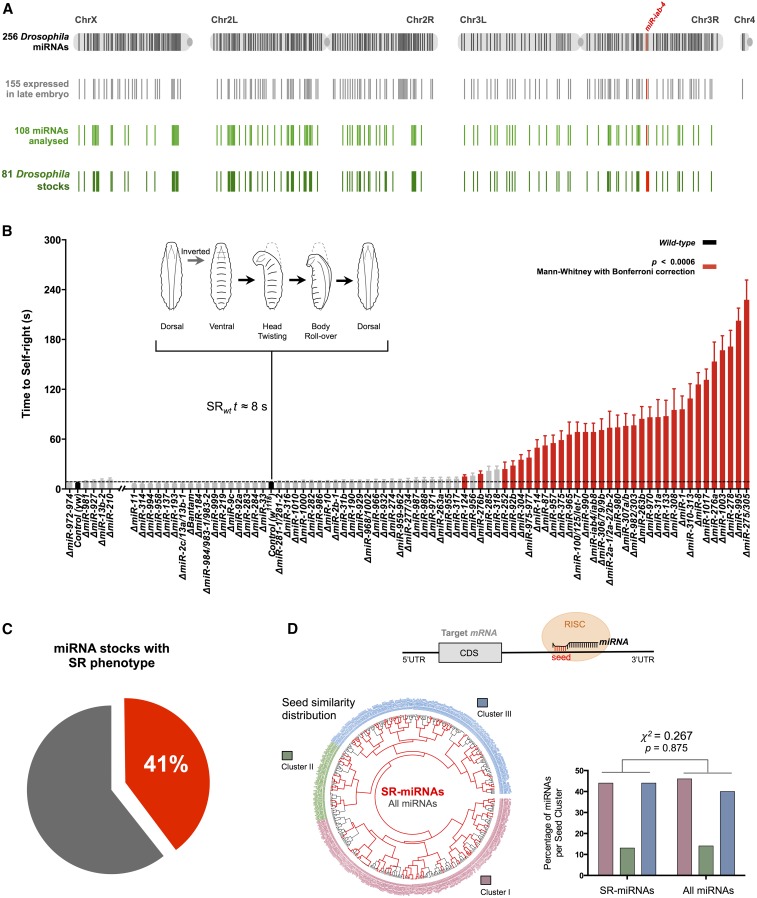
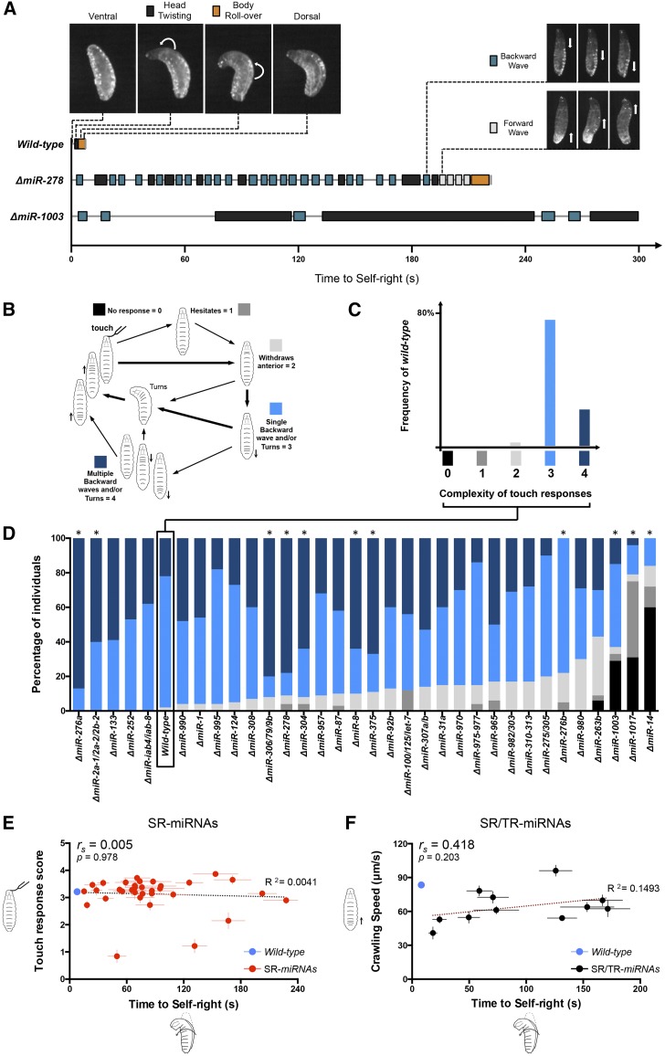
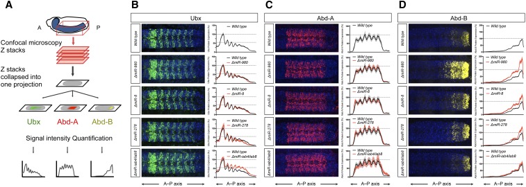
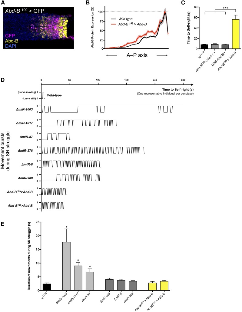
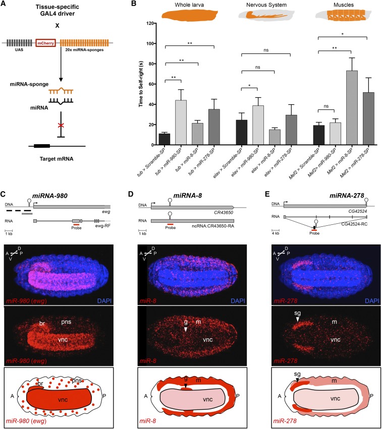
The effects of microRNA (miRNA) regulation on the genetic programs underlying behavior remain largely unexplored. Despite this, recent work in Drosophila shows that mutation of a single miRNA locus (miR-iab4/iab8) affects the capacity of the larva to correct its orientation if turned upside down (self-righting, SR), suggesting that other miRNAs might also be involved in behavioral control. Here we explore this possibility, studying early larval SR behavior in a collection of 81 Drosophila miRNA mutants covering almost the entire miRNA complement of the late embryo. Unexpectedly, we observe that >40% of all miRNAs tested significantly affect SR time, revealing pervasive behavioral effects of miRNA regulation in the early larva. Detailed analyses of those miRNAs affecting SR behavior (SR-miRNAs) show that individual miRNAs can affect movement in different ways, suggesting that specific molecular and cellular elements are affected by individual miRNA mutations. Furthermore, gene expression analysis shows that the Hox gene Abdominal-B (Abd-B) represents one of the targets deregulated by several SR-miRNAs. Our work thus reveals pervasive effects of miRNA regulation on a complex innate behavior in Drosophila and suggests that miRNAs may be core components of the genetic programs underlying behavioral control in other animals too.

Keywords: CNS; Drosophila; Hox genes; behavior; miRNA regulation; microRNA; nervous system.


Figures

Figure 1
Figure 1
Pervasive role of miRNAs in self-righting behavior. (A) Graphic representation of the miRNA precursor sequences along the four Drosophila chromosomes. The total 256 miRNA precursor sequences from the latest miRBase version (miRBase 21) (Kozomara and Griffiths-Jones 2014) are represented in black lines. The 155 miRNAs expressed in late embryos are represented in gray lines (data accessible at National Center for Biotechnology Information GEO database (Barrett et al. 2013) accession no. GSM364902; 12- to 24-hr Drosophila embryos). Represented in green are the 108 individual miRNA precursors (light green lines) included in the 81 mutant stocks analyzed (dark green lines) (File S1 and File S5). In red is the miR-iab-4 that had been previously described to disrupt SR (Picao-Osorio et al. 2015). (B) Quantification of the time required for successful completion of self-righting (mean ± SEM; with an average of 29 larvae per genotype). The two different miRNA mutant genetic backgrounds—yw and w1118—were compared with the respective controls (in black) and are separated in two groups: yw and five mutants and w1118 and 76 mutants. The mutants showing SR delay with statistical significance of P ≤ 0.0006 (Mann–Whitney U-test with Bonferroni correction) are depicted in red. Representation of the sequential movements of SR is depicted above: when placed in an inverted position (ventral up), larvae twist their heads and roll their bodies onto their ventral surface (dorsal up). This sequence takes an average of 8 sec in control (wt) larvae. (C) Pie chart of the percentage of miRNA mutant stocks (33 out of 81) showing a statistically significant delay in SR time. (D, top) Diagram illustrating the miRNA-mediated downregulation of gene expression through association with the RNA-induced silencing complex (RISC) and seed pairing with a 3′-UTR target. (D) Hierarchical clustering of the D. melanogaster miRNA seed sequence complement (positions 2–7, gray). The distribution of SR-miRNA seed sequences (red) within the Drosophila miRNA seed-sequence space (black) is identical to the distribution of all Drosophila mature miRNAs (right, Pearson’s χ2-test = 0.267, P = 0.875).
Figure 2
Figure 2
Diversity of miRNA effects on SR. (A, top left) Representative wild-type SR sequence in a single larva with two main phases: head twisting (black) and body roll-over (orange). (A, top right) Representation of other SR-miRNA movements while in an inverted position during SR struggles: backward (blue) and forward (light gray) waves. (A, bottom) Representative examples of SR sequences in single miRNA mutant larvae, depicting the occurrence of different movements and their frequency and duration. miR-278 mutant larvae are active, frequently alternating between different movements (e.g., forward and backward waves) until they are able to roll over their bodies. ΔmiR-1003 larvae generally take a longer time performing each SR phase. More SR-miRNA mutant examples are described in Figure 4D. (B) Representation of the different responses to touch in the anterior region and respective scores (0–4) based on Kernan et al. (1994) (TR). Insensitivity to touch is shown in black (score 0). Simpler and more complex responses are represented in shades of gray and blue, respectively, and are translated in scores ranging from 1 to 4. (C) Frequency of the different responses to touch in wild-type larvae. Simpler responses (shades of gray) are extremely rare. While around 20% of the individuals show more complex responses (with multiple backward waves), the most common response (∼80% of the cases) is obstacle avoidance through turning or performance of a single backward wave followed by turning. (D) Percentage of the different TR scores in each miRNA mutant (N = 15–33 larvae per genotype). * represents significant deviations from the wild-type responses (w1118, black box) with P < 0.0015 (Mann–Whitney U-test with Bonferroni correction). (E) Correlation between TR scores (y-axis) and time to SR (x-axis) of all 33 SR-miRNAs. The wild-type genotype is depicted by a blue circle, the SR-miRNAs by red circles, and linear regression in dotted black line (R2 = 0.0041). The Spearman coefficient (rs) and P-value are shown. There is no significant correlation between TR and the SR delay (rs = 0.005; P = 0.978). (F) Correlation between crawling speed (micrometers per second) of freely exploratory larvae behavior (y-axis) (mean ± SEM; with an average of 17 larvae per genotype) and time to self-right (x-axis) of the 11 mutants showing both SR and TR phenotypes (SR/TR-miRNAs, black circles). Wild type is depicted by a blue circle. Linear regression line in dark red (R2 = 0.1493). The Spearman coefficient (rs) and P-value are shown. There is no significant correlation between crawling speed and the SR delay (rs = 0.4182; P = 0.203).
Figure 3
Figure 3
SR-miRNAs control Hox gene expression. (A) Schematic representation of Hox protein expression analysis along the A–P axis (see Materials and Methods). Immunostained whole-mounted embryos for the three BX-C proteins were imaged using confocal microscopy. Confocal stacks of the ventral nerve cord of each specimen were collapsed into one projection and levels of expression along the A–P axis quantified. (B–D, left) Embryonic protein expression of Ubx (B, green), Abd-A (C, red), and Abd-B (D, yellow) in the ventral nerve cord of wild-type and mutants for miR-980, miR-8, miR-278, and miR-iab-4/iab-8 at late 16 stage. (B–D, right) Profile quantification along the A–P axis for the three Hox proteins in the wild type (mean in black line and SEM in gray) and in the miRNA mutants (mean in red line and SEM in lighter red). (B) Only ΔmiR-iab-4/iab-8 shows differences in Ubx protein expression (as previously described by Bender 2008; Thomsen et al. 2010; Picao-Osorio et al. 2015). (C) No significant expression change was observed in Abd-A protein in the four miRNA mutants. (D) Significant expression change in Abd-B protein for miR-980, miR-8, and miR-278 mutants. N = 10 embryos per genotype for each immunostaining. DAPI is in blue. Anterior is to the left.
Figure 4
Figure 4
Overexpression of Abd-B disrupts SR behavior. (A) Expression pattern of Abd-B199-GAL4 driver (GFP, magenta) with respect to the endogenous pattern of Abd-B protein expression (yellow) in dissected embryonic ventral nerve cord. DAPI is in blue and anterior is to the left. (B) Quantification of Abd-B expression profile along the A–P axis in dissected embryonic nerve cords of wild-type (w1118, mean in black and SEM in gray) and Abd-B overexpression (Abd-B199 > Abd-B, mean in magenta and SEM in light magenta) (N = 9 embryos per genotype). (C) Significant delay in time to SR in larvae overexpressing Abd-B (Abd-B199 > Abd-B, yellow bars) in comparison with wild-type (w1118) and parental lines (Abd-B199-GAL4/+, light gray bar, and UAS-Abd-B/+ in dark gray) (mean ± SEM; an average of 20 larvae per genotype were analyzed; Mann–Whitney U-test with Bonferroni correction, *** P < 0.001). (D) Representative examples of activity patterns of SR struggle of single miRNA mutant larvae and Abd-B overexpressing larvae. During the SR routine, we assigned a value of 1 if the larva performed any of the movements mentioned in Figure 2A, and 0 when it remained still. Mutants miR-1003, miR-1017, and miR-87 represent examples of SR-miRNA mutants that take a longer to perform each SR phase. In contrast, mutant larvae for miR-278, miR-8, and miR-980 are examples of active larvae that frequently alternate between different movements (e.g., forward and backward waves) until they are able to roll over their bodies. The characteristic SR struggle of Abd-B overexpressing larvae (Abd-BLDN > Abd-B and Abd-B199 > Abd-B) was comparable to miR-278, miR-8, and miR-980. (E) Quantification of duration of each movement during SR behavior. SR-miRNA mutants miR-1003, miR-1017, and miR-87 (light gray bars) showed longer periods on each SR movement compared to the wild type (w1118, black bar). Conversely, SR-miRNA mutants miR-278, miR-8, and miR-980 (dark gray bars) and larvae overexpressing Abd-B (yellow bars) showed similar duration on each SR movement compared to the wild type (mean ± SEM; N = 4 larvae; Mann–Whitney U-test, * P < 0.05).
Figure 5
Figure 5
SR-miRNAs disrupt SR behavior through action in different tissues. (A) Schematic representation of miRNA-sponges (miR-SP) mode of action (based on Fulga et al. 2015): tissue-specific expression of mCherry constructs containing 20 miRNA-specific binding sites in the 3′-UTR. miRNAs will bind to the miR-SP, reducing the available RISC–miRNA complex that represses endogenous mRNA targets. (B) SR behavior in tissue-specific knockdown of miRNAs. miR-SPs were expressed in all larval tissues (tubulin-Gal4, left), nervous system (elavc155-Gal4, center), and muscle (Mef2-Gal4, right). Scramble-SP (UAS-sponge with a scrambled sequence; black bars) was used as control. miR-980-SP (light gray bars), miR-8-SP (gray bars), and miR-278-SP (dark gray bars) were used to knock down the miRNAs miR-980, miR-8, and miR-278, respectively. All three miR-SPs show a statistically significant delay in SR time compared with Scramble-SP when expressed ubiquitously (left, tub > miR-SP). Only miR-980-SP significantly recapitulated this SR delay when expressed exclusively in the nervous system (middle, elav > miR-980-SP), while miR-8-SP and miR-278-SP disrupted SR behavior when expressed in the muscle (right, Mef2 > miR-8-SP and Mef2 > miR-278-SP). Bars represent mean ± SEM; an average of 33 larvae per genotype were analyzed; Mann–Whitney U-test; ns, nonsignificant, * P < 0.05, ** P < 0.01). Additionally see Figure S6 in File S4 for parental line controls. (C–E) Embryonic expression of miR-980, miR-8, and miR-278. (C–E, top) Schematic representation of miR-980, miR-8, and miR-278 loci. FISH RNA probes generated to detect the primary miRNA (pri-miRNA) transcripts are shown in red rectangles. Note that the full transcription unit of miR-980 is unknown. None of the several probes designed and tested to detect the expression of the primary transcript of miR-980 were successful (black rectangles represent probes used for conventional FISH and the black barred rectangle indicates the region of the 44 probes used for single-molecule FISH; data not shown). Given that the transcription start site of erect wing (ewg) is ∼500 bp downstream of miR-980, we used a probe targeting an exon present in all ewg mRNA isoforms (red rectangle) as a proxy for miR-980 spatial expression. miR-8 is located in the CR43650 long noncoding RNA, while miR-278 is coded within the 3′-UTR of CG42524 mRNA isoform C. (C–E, middle) Spatial expression of pri-miR-980 (ewg), pri-miR-8, and pri-miR-278 obtained by RNA FISH using the red probes in the top panel. Whole-mounted late 16-stage embryos were imaged using confocal microscopy. (C) pri-miR-980 (ewg) is expressed predominantly in the CNS and with punctual expression in PNS. (D) pri-miR-8 is highly expressed in the muscle, in some neurons along the ventral nerve cord, and in the anterior gut. (E) pri-miR-278 is strongly expressed in the salivary glands and in a few scattered muscle and CNS cells. DAPI is in blue. Anterior is to the left. br, brain; g, gut; m, muscle; pns, peripheral nervous system; sg, salivary glangs; vnc, ventral nerve cord. (C–E, bottom) Diagrams showing the expression patterns of miR-980, miR-8, and miR-278 at late embryogenesis. Dark red represents high expression and light red depicts expression in small groups of cells.

Similar articles

Cited by

References

    1. Aboobaker A. A., Tomancak P., Patel N., Rubin G. M., Lai E. C., 2005. Drosophila microRNAs exhibit diverse spatial expression patterns during embryonic development. Proc. Natl. Acad. Sci. USA 102: 18017–18022. - PMC - PubMed
    1. Allada R., White N. E., So W. V., Hall J. C., Rosbash M., 1998. A mutant Drosophila homolog of mammalian Clock disrupts circadian rhythms and transcription of period and timeless. Cell 93: 791–804. - PubMed
    1. Alonso C. R., 2012. A complex “mRNA degradation code” controls gene expression during animal development. Trends Genet. 28: 78–88. - PubMed
    1. Armstrong J. D., Texada M. J., Munjaal R., Baker D. A., Beckingham K. M., 2006. Gravitaxis in Drosophila melanogaster: a forward genetic screen. Genes Brain Behav. 5: 222–239. - PubMed
    1. Baek D., Villén J., Shin C., Camargo F. D., Gygi S. P., et al. , 2008. The impact of microRNAs on protein output. Nature 455: 64–71. - PMC - PubMed

Publication types

LinkOut - more resources