. 2017 Jul 1;313(1):C42-C53.
doi: 10.1152/ajpcell.00244.2016. Epub 2017 May 3.

ENaC activity is regulated by calpain-2 proteolysis of MARCKS proteins

Affiliations

ENaC activity is regulated by calpain-2 proteolysis of MARCKS proteins

Darrice S Montgomery et al. Am J Physiol Cell Physiol. .

Abstract

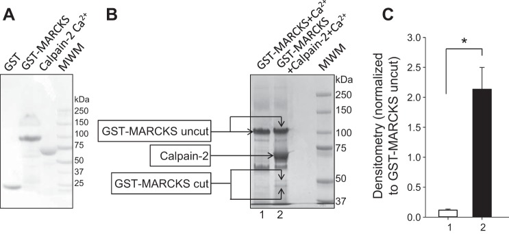
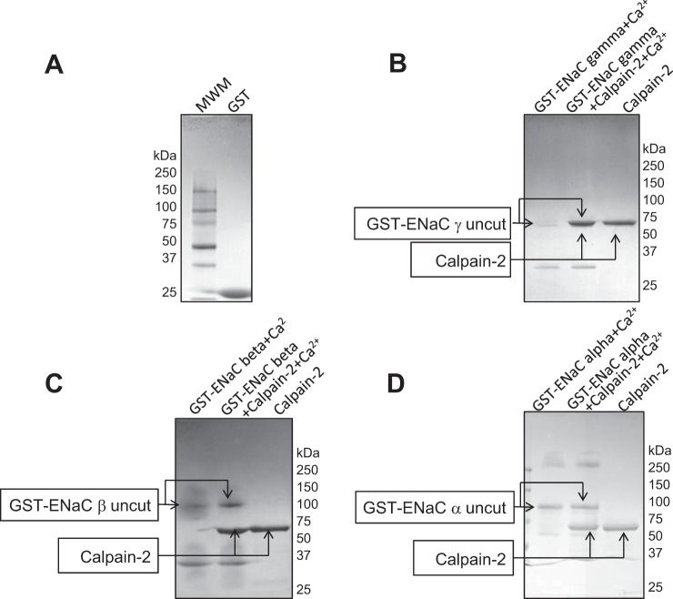
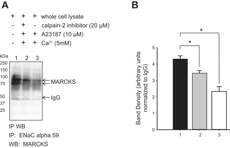
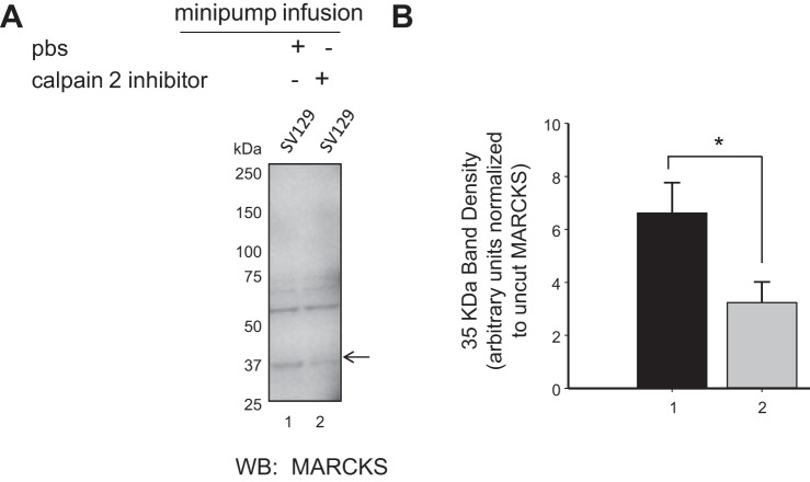
We previously demonstrated a role for the myristoylated alanine-rich C kinase substrate (MARCKS) to serve as an adaptor protein in the anionic phospholipid phosphate-dependent regulation of the epithelial sodium channel (ENaC). Both MARCKS and ENaC are regulated by proteolysis. Calpains are a family of ubiquitously expressed intracellular Ca2+-dependent cysteine proteases involved in signal transduction. Here we examine the role of calpain-2 in regulating MARCKS and ENaC in cultured renal epithelial cells and in the mouse kidney. Using recombinant fusion proteins, we show that MARCKS, but not the ENaC subunits, are a substrate of calpain-2 in the presence of Ca2+ Pharmacological inhibition of calpain-2 alters MARCKS protein expression in light-density sucrose gradient fractions from cell lysates of mouse cortical collecting duct cells. Calpain-dependent cleaved products of MARCKS are detectable in cultured renal cells. Ca2+ mobilization and calpain-2 inhibition decrease the association between ENaC and MARCKS. The inhibition of calpain-2 reduces ENaC activity as demonstrated by single-channel patch-clamp recordings and transepithelial current measurements. These results suggest that calpain-2 proteolysis of MARCKS promotes its interaction with lipids and ENaC at the plasma membrane to allow for the phosphatidylinositol 4,5-bisphosphate (PIP2)-dependent regulation of ENaC activity in the kidney.

Keywords: calcium; calpain; epithelial sodium channel; myristoylated alanine-rich C kinase substrate; protein kinase C; proteolysis.


Figures

Fig. 1.
Fig. 1.
SDS-PAGE and Coomassie staining analysis of MARCKS cleavage by calpain-2. A: recombinant GST and GST-MARCKS expressed and purified from BL21 cells are shown to have an electrophoretic mobility of 26 and 100 kDa, respectively. Active calpain-2 purified from porcine kidney is shown to have an electrophoretic mobility of 70 kDa in the presence of Ca2+. B: representative Coomassie gel showing the cleavage of GST-MARCKS after incubation with active calpain-2 in the presence of CaCl2 or with CaCl2 only for 1 h at 37°C. Lower-molecular weight bands above and below 50 kDa are shown in the lane with GST-MARCKS incubated with active calpain-2 in the presence of Ca2+ but absent in the lane with GST-MARCKS and Ca2+ alone. MWM, molecular weight markers. C: densitometric analysis of calpain proteolysis of MARCKS as shown in B. The data are representative of three independent experiments (n = 3). Data are represented as means ± SE. *P < 0.001 by t-test.
Fig. 2.
Fig. 2.
SDS-PAGE and Coomassie staining analysis of ENaC cleavage by calpain-2. A: recombinant GST expressed and purified from BL21 cells are shown to have an electrophoretic mobility of 26 kDa. MWM, molecular weight markers. Recombinant GST-ENaCγ (B), GST-ENaCβ (C), and GST-ENaCα (D) fusion proteins purified from bacterial inclusion bodies were incubated with active calpain-2 in the presence of CaCl2 or with CaCl2 only for 1 h at 37°C. Lower-molecular weight bands corresponding to cleaved products from calpain-2 proteolysis are absent, and there is no appreciable difference between any of the three ENaC subunits incubated with or without calpain-2 in the presence of CaCl2. Active calpain-2 purified from porcine kidney was loaded separately as a control.
Fig. 3.
Fig. 3.
Amiloride-sensitive transepithelial current measurements examining the effect of active calpain-2 or calpain-2 inhibition in mpkCCD cells. A: active calpain-2 from porcine kidney was applied to the apical side of confluent mpkCCD cell monolayers. Transepithelial voltages and resistances were recorded, and equivalent transepithelial current was calculated using Ohm’s law. There was no change in current over time between cells treated with calpain-2 and untreated cells. Amiloride (0.5 µM) was applied to the apical side of the cells at the end of the experiment to show that current from any amiloride-insensitive nonselective cation channel was negligible. Current was calculated from three separate inserts (n = 3). B: effect of calpain-2 inhibition on amiloride-sensitive transepithelial current in mpkCCD cells by epithelial voltohmmeter measurements. Calpain-2 inhibitor (20 μM) or cytochalasin D (1.5 μM) as a control was applied to the apical side of confluent mpkCCD cell monolayers. Transepithelial current was calculated from transepithelial voltage and resistance measurements. At the end of the experiment, amiloride (0.5 µM) was added to show that the measured current was from amiloride-sensitive highly selective cation channels. Current was calculated from three separate inserts (n = 3) for each group. Data are represented as means ± SE. *P < 0.05 compared with untreated cells.
Fig. 4.
Fig. 4.
Cell-attached single-channel patch-clamp recordings examining the activity of ENaC in Xenopus 2F3 cells treated with or without the calpain-2 inhibitor A6060. A: representative recordings from Xenopus 2F3 cells after pretreating the apical and basolateral sides of the cells with or without 20 μM calpain-2 inhibitor for 6 h. All recordings were made at 0-mV holding potential. B: summary bar graph of ENaC activity (NPo) in the control and calpain-2 inhibitor groups. ENaC activity was substantially reduced in cells treated with the calpain-2 inhibitor compared with cells treated with vehicle alone. A total of 20 patches (n = 20) were obtained and analyzed for the control group, and 13 patches (n = 13) were obtained and analyzed for the group of cells treated with the calpain-2 inhibitor. Data are represented as means ± SE. ***Significant difference (P < 0.01) between the two groups. Current-voltage (I–V) relationship for the control group (C) and the calpain inhibitor group (D). All holding potential (Vpip) values are indicated as −Vpip. The conductance from control cells was 5.52 ± 0.20 pS, whereas the conductance from the calpain-inhibited cells was 5.34 ± 0.22.
Fig. 5.
Fig. 5.
Sucrose density gradient and Western blot analysis of calpain-2 inhibition on MARCKS protein expression in mpkCCD cells. A: representative Western blot (WB) showing MARCKS expression in various fractions including light-density sucrose gradient fractions. B: representative Western blot showing a substantial decrease in MARCKS protein expression in light-density sucrose gradient fractions after treating mpkCCD cells with the pharmacological inhibitor of calpain-2 A6060 compared with mock (vehicle) treatment. Flotillin was used as a marker of lipid rafts in A and B. C: densitometric analysis of the Western blot data (n = 3) presented in A. Data are represented as means ± SE. *P < 0.05, **P < 0.01.
Fig. 6.
Fig. 6.
Lipid binding analysis of calpain-2 and the effect of calpain-2 proteolysis of MARCKS on the direct binding with lipids. A: indirect overlay binding assay probing for interactions between recombinant calpain-2 and 15 different lipids spotted on a nitrocellulose membrane (see materials and methods). The membrane was probed with specific anti-calpain-2 rabbit polyclonal antibody followed by incubation with an anti-rabbit secondary antibody. There was no appreciable binding of any of the lipids and calpain-2 after exposure of the membrane. Representative direct overlay binding assay probing for interactions between recombinant GST-MARCKS immobilized on a Sepharose support after incubation with calpain-2 (B) or without calpain-2 (C) in the presence of CaCl2 before eluting the bound GST-MARCKS fusion protein with reduced glutathione. A peroxidase-conjugated anti-GST antibody was used to probe for binding between calpain-cleaved MARCKS or uncleaved MARCKS and 15 different lipids. D: direct overlay binding assay in which recombinant GST, as a negative control, was incubated with a nitrocellulose membrane containing 15 different lipids before probing the membrane with a peroxidase-conjugated anti-GST antibody. E: identification of the 15 lipids spotted on the nitrocellulose membrane. F: densitometric analysis of the representative overlay binding assay data presented in B and C. Summary bar graph showing differences between calpain-cleaved GST-MARCKS (n = 3) and uncleaved GST-MARCKS (n = 3) in the binding of 3,4-PIP2, 4,5-PIP2, and 3,4,5-PIP3. Arrows indicate differences in binding between GST-MARCKS cleaved by calpain-2 (B) and uncleaved GST-MARCKS (C). Units are equivalent for all bars in graph. Data are represented as means ± SD. *P < 0.001 determined by Kruskal-Wallis ANOVA on ranks with Student-Neuman-Keuls posttest.
Fig. 7.
Fig. 7.
Immunoprecipitation and Western blot analysis showing the effect of Ca2+ mobilization and calpain-2 inhibition on the association between ENaCα and MARCKS. A: representative immunoprecipitation (IP) Western blot showing the association between ENaCα and MARCKS in response to intracellular calcium mobilization and/or calpain-2 inhibition. Xenopus 2F3 cells were incubated with Ca2+, the Ca2+ ionophore A23187, and/or calpain-2 inhibitor for 6 h before harvesting the cells for protein. ENaCα was immunoprecipitated from the whole cell lysate using specific antibody (ENaCα 59). The blot was probed for MARCKS using specific antibody. The association between ENaCα and MARCKS was decreased in cells treated with 5 mM Ca2+ in the presence of the Ca2+ ionophore A23187 and in cells treated with the calpain-2 inhibitor A6060. B: densitometric analysis of the immunoreactive bands in A. The data are representative of three independent experiments (n = 3). Arbitrary units represent a relative unit of measurement of the band intensities between groups determined with ImageJ. Units are equivalent for all bars in graph. Data are represented as means ± SE. *P < 0.05 determined by Kruskal-Wallis ANOVA on ranks with Student-Neuman-Keuls posttest.
Fig. 8.
Fig. 8.
Calpain-mediated MARCKS cleavage in native renal cells. Mouse mpkCCDc14 cells were transfected with a Flag-tagged MARCKS construct. Confluent cells were divided into three groups: untreated (black bars), treated with 100 μM hydrogen peroxide for 30 min to activate calpain, or treated with 100 μM hydrogen peroxide (30 min) plus 50 nM calpain inhibitor acetyl-calpastatin for 2–12 h. We then blotted for Flag or MARCKS. With Flag we observed two bands, which we presume are uncleaved and partially cleaved MARCKS (band a, uncleaved; band #, cleaved). In the presence of inhibitor the uncleaved MARCKS increases significantly, and the partially cleaved MARCKS decreases [data from 6 blots (n = 6) determined by Kruskal-Wallis ANOVA on ranks with Student-Neuman-Keuls posttest]. After stripping the Flag blots, we blotted for MARCKS and observed two bands: one that corresponds to uncleaved MARCKS (band b) and a lower-molecular-weight cleaved product (band c) that increases after exposure to peroxide and is reduced in the presence of inhibitor (data from 6 blots determined by Kruskal-Wallis ANOVA on ranks with Student-Neuman-Keuls posttest). Units are equivalent for all bars in graphs. Data are represented as means ± SE. *P < 0.05.
Fig. 9.
Fig. 9.
Western blot analysis of MARCKS protein expression in native kidney after infusion of the calpain-2 inhibitor A6060 in mice. Osmotic minipumps were used to infuse the calpain-2 inhibitor A6060 or 50% DMSO in sterile saline in SV129 mice at a rate of 0.5 μl/h for 7 days. A: representative Western blot of three independent experiments probing for MARCKS protein expression shows immunoreactive bands at 75 (doublet), 60, and 37 kDa. The density of the 37-kDa cleaved form of MARCKS was less in lysates from kidneys infused with calpain-2 inhibitor compared with PBS in SV129 mice. B: densitometric analysis of the immunoreactive band at 37 kDa in A representative of three independent experiments (n = 3). Data are represented as means ± SE. *P < 0.05 determined by t-test.
Fig. 10.
Fig. 10.
Proposed model describing the role of calpain-2 in the MARCKS-mediated PIP2-dependent regulation of ENaC in the kidney. A: in the presence of active calpain-2 a region downstream of the effector domain of MARCKS is cleaved. Calpain-2 proteolysis of MARCKS prevents PKCα from accessing and phosphorylating serine residues within the effector domain resulting in constitutive or increased MARCKS association with the membrane. At the apical plasma membrane, MARCKS sequesters acidic phospholipid phosphates (e.g., PIP2) and presents them to ENaC. These phospholipid phosphates positively regulate the gating of ENaC and maintain the channel in an open conformation. B: conversely, in the absence of active calpain-2, MARCKS is not cleaved, and the carboxy-terminal tail potentiates the access of PKCα to the effector domain of MARCKS. PKCα phosphorylates serine residues within the effector domain of MARCKS and causes the protein to lose affinity for the membrane and translocate to the cytoplasm. In turn, the interaction between PIP2 and ENaC is reduced because both molecules are rare and do not associate by random diffusion alone. PIP2 is shown to be randomly dispersed within the inner leaflet of the apical plasma membrane instead of being in close proximity to ENaC after MARCKS is phosphorylated by PKC and translocated to the cytoplasm. C-terminus, COOH terminus.

Similar articles

Cited by

References

    1. Aderem A. The MARCKS brothers: a family of protein kinase C substrates. Cell 71: 713–716, 1992. doi:10.1016/0092-8674(92)90546-O. - DOI - PubMed
    1. Aki T, Yoshida K, Fujimiya T. Phosphoinositide 3-kinase accelerates calpain-dependent proteolysis of fodrin during hypoxic cell death. J Biochem 132: 921–926, 2002. doi:10.1093/oxfordjournals.jbchem.a003305. - DOI - PubMed
    1. Alli AA, Bao HF, Alli AA, Aldrugh Y, Song JZ, Ma HP, Yu L, Al-Khalili O, Eaton DC. Phosphatidylinositol phosphate-dependent regulation of Xenopus ENaC by MARCKS protein. Am J Physiol Renal Physiol 303: F800–F811, 2012. doi:10.1152/ajprenal.00703.2011. - DOI - PMC - PubMed
    1. Alli AA, Bao HF, Liu BC, Yu L, Aldrugh S, Montgomery DS, Ma HP, Eaton DC. Calmodulin and CaMKII modulate ENaC activity by regulating the association of MARCKS and the cytoskeleton with the apical membrane. Am J Physiol Renal Physiol 309: F456–F463, 2015. doi:10.1152/ajprenal.00631.2014. - DOI - PMC - PubMed
    1. Alli AA, Gower WR Jr. Molecular approaches to examine the phosphorylation state of the C type natriuretic peptide receptor. J Cell Biochem 110: 985–994, 2010. doi:10.1002/jcb.22612. - DOI - PubMed

MeSH terms

LinkOut - more resources