. 2017 Jul 7;28(14):1975-1983.
doi: 10.1091/mbc.E17-03-0171. Epub 2017 May 3.

Increased lateral microtubule contact at the cell cortex is sufficient to drive mammalian spindle elongation

Affiliations

Increased lateral microtubule contact at the cell cortex is sufficient to drive mammalian spindle elongation

Joshua Guild et al. Mol Biol Cell. .

Abstract

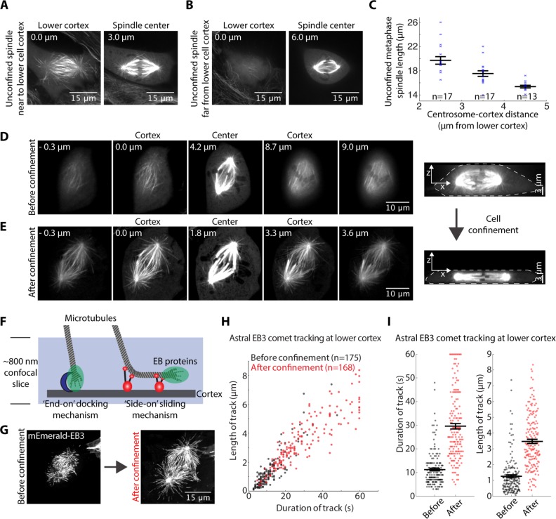
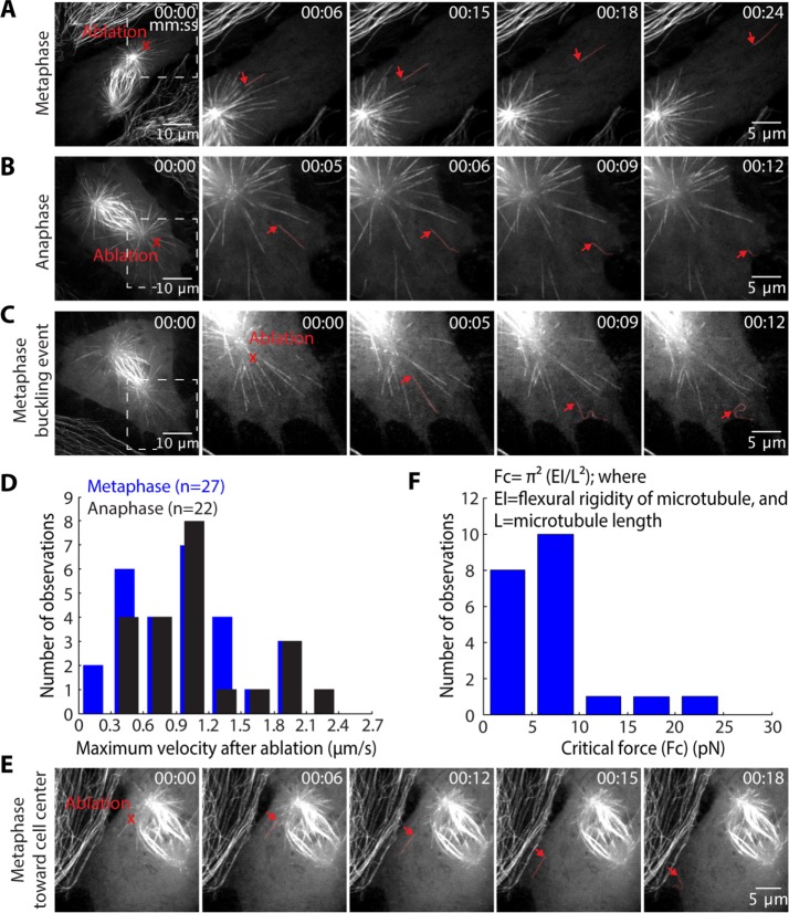
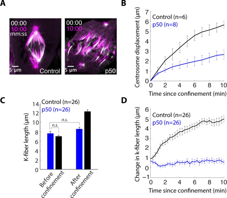
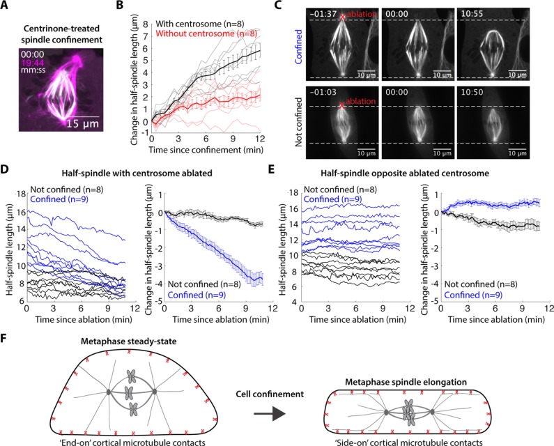
The spindle is a dynamic structure that changes its architecture and size in response to biochemical and physical cues. For example, a simple physical change, cell confinement, can trigger centrosome separation and increase spindle steady-state length at metaphase. How this occurs is not understood, and is the question we pose here. We find that metaphase and anaphase spindles elongate at the same rate when confined, suggesting that similar elongation forces can be generated independent of biochemical and spindle structural differences. Furthermore, this elongation does not require bipolar spindle architecture or dynamic microtubules. Rather, confinement increases numbers of astral microtubules laterally contacting the cortex, shifting contact geometry from "end-on" to "side-on." Astral microtubules engage cortically anchored motors along their length, as demonstrated by outward sliding and buckling after ablation-mediated release from the centrosome. We show that dynein is required for confinement-induced spindle elongation, and both chemical and physical centrosome removal demonstrate that astral microtubules are required for such spindle elongation and its maintenance. Together the data suggest that promoting lateral cortex-microtubule contacts increases dynein-mediated force generation and is sufficient to drive spindle elongation. More broadly, changes in microtubule-to-cortex contact geometry could offer a mechanism for translating changes in cell shape into dramatic intracellular remodeling.


Figures

FIGURE 1:
FIGURE 1:
Metaphase, anaphase, monopolar, and Taxol-stabilized spindles elongate at similar rates when confined. (A) Schematic diagram of PDMS-based cell confinement. (B, C) Confocal images of representative examples of (B) confinement-induced metaphase spindle elongation and (C) anaphase B spindle elongation in a confined cell. (D) Metaphase and anaphase spindle length following confinement. (E) Mean ± SEM (thick line) and individual traces (thin lines) of change in spindle length for metaphase and anaphase spindles following confinement. (F) Representative example of confinement-induced (STLC-induced, 10 µM) monopolar spindle elongation. (G) Schematic and (H) mean ± SEM (thick line) and individual traces (thin lines) of path length of centrosome movement following confinement in metaphase, anaphase, and monopolar spindles. (I) Representative example of confinement-induced Taxol-treated (10 µM) metaphase spindle elongation. (J) Mean ± SEM (thick line) and individual traces (thin lines) of change in spindle length for metaphase and Taxol-treated metaphase spindles following confinement. (K) Example sister kinetochore pair (mCherry-CenpC) demonstrating that k-fibers (GFP-tubulin) can fall off kinetochores to allow spindle elongation in Taxol. For B, C, F, and I, phase-contrast and GFP-tubulin images are merged. For all data, PtK2 GFP-tubulin cells were captured by confocal imaging and confinement occurs at t = 0 and persists thereafter.
FIGURE 2:
FIGURE 2:
Confinement promotes lateral microtubule growth at the cell cortex. (A, B) Confocal images of the lower cortex (0 μm) and spindle center (distance from cortex noted in micrometers) in unconfined PtK2 GFP-tubulin metaphase cells with (A) a “long” spindle nearer the cortex and (B) a “short” spindle farther from the cortex. (C) Unconfined metaphase spindle length (mean ± SEM) correlates with distance of centrosomes to the lower cell cortex. (D, E) Confocal z-stacks of the same PtK2 GFP-tubulin metaphase spindle (D) before and (E) after confinement (with lower cortex at 0 μm, and upper cortex distance noted). (F) Schematic demonstrating alternative force-generating mechanisms and microtubule contact geometries at the cell cortex during mitosis. (G) mEmerald-EB3 maximum-intensity projection (from confocal images) over time at the lower cell cortex for a 1 min period before and after metaphase cell confinement. (H) Length and duration of EB3 comet tracks at the lower cell cortex (n = 4 cells analyzed both before and shortly after confinement). (I) Duration and length of comet tracks (mean ± SEM) from H are plotted separately to display statistical significance.
FIGURE 3:
FIGURE 3:
Force is applied along the length of astral microtubules at both metaphase and anaphase. Confocal images of (A) metaphase and (B) anaphase unconfined PtK2 GFP-tubulin cells in which laser ablation (red “X”) was used to sever astral microtubules near the centrosome at the lower cell cortex. Severed astral microtubules (red) moved away from the spindle. (C) Severed microtubules (red) frequently buckled against the cortex. (D) Histogram of the maximum speed reached by each severed microtubule at metaphase and anaphase (n = number of cuts). (E) In a metaphase cell, an astral microtubule extending into the spindle is pulled toward the cell edge upon detachment from the spindle (ablation at red “X”; arrow points to new minus end). (F) Histogram of calculated critical forces required for observed buckling events at both metaphase and anaphase.
FIGURE 4:
FIGURE 4:
Dynein is required for confinement-induced spindle elongation. (A) Confocal images of PtK2 GFP-tubulin cell immediately following confinement (white) and 10 min after confinement (magenta) in a control cell and a cell overexpressing p50 to inhibit dynein. (B) Centrosome displacement (mean ± SEM, n = number of centrosomes) from the original position following confinement in control and p50-overexpressing cells. (C) K-fiber length (mean ± SEM, n = number of k-fibers) before confinement and 12 min after confinement in p50 overexpression and control cells. (D) Change in k-fiber length (mean ± SEM, n = # k-fibers) following confinement in p50-overexpressing and control cells.
FIGURE 5:
FIGURE 5:
Astral microtubules are required for confinement-induced spindle elongation. (A) Confocal images of a centrinone-treated (125 nM) PtK2 GFP-tubulin cell immediately following (white) and almost 20 min after confinement (magenta). (B) Half-spindle elongation (mean ± SEM) following confinement of centrinone-treated cells. Confinement occurs at t = 0. (C) Time-lapse confocal images of PtK2 GFP-tubulin cells demonstrating the representative asymmetric response to centrosome ablation (at t = 0, red “X”) in a cell confined 15 min before ablation (top) and an unconfined (bottom) cell. (D, E) Half-spindle length and changes in half-spindle length (mean ± SEM) following ablation (t = 0) in the (D) half-spindle with centrosome ablated at t = 0 or (E) half-spindle opposite the ablated centrosome. (F) Model schematic: confinement increases sliding forces generated by dynein at the cell cortex on astral microtubules, and these forces are both sufficient to induce spindle elongation and necessary for maintenance of the elongated state.

References

    1. Adames NR, Cooper JA. Microtubule interactions with the cell cortex causing nuclear movements in Saccharomyces cerevisiae. J Cell Biol. 2000;149:863–874. - PMC - PubMed
    1. Aist JR, Liang H, Berns MW. Astral and spindle forces in PtK2 cells during anaphase B: a laser microbeam study. J Cell Sci. 1993;104:1207–1216. - PubMed
    1. Brennan IM, Peters U, Kapoor TM, Straight AF. Polo-like kinase controls vertebrate spindle elongation and cytokinesis. PLoS One. 2007;2:e409. - PMC - PubMed
    1. Canman JC, Cameron LA, Maddox PS, Straight A, Tirnauer JS, Mitchison TJ, Fang G, Kapoor TM, Salmon ED. Determining the position of the cell division plane. Nature. 2003;424:1074–1078. - PubMed
    1. Collins ES, Balchand SK, Faraci JL, Wadsworth P, Lee WL. Cell cycle-regulated cortical dynein/dynactin promotes symmetric cell division by differential pole motion in anaphase. Mol Biol Cell. 2012;23:3380–3390. - PMC - PubMed