

Rapid and specific detection of Asian- and African-lineage Zika viruses
- PMID: 28469032
- PMCID: PMC5654541
- DOI: 10.1126/scitranslmed.aag0538
Rapid and specific detection of Asian- and African-lineage Zika viruses
Abstract
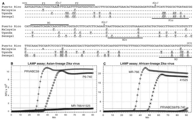
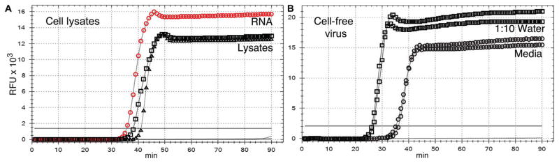
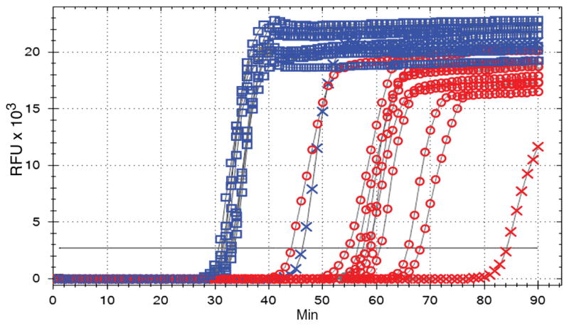
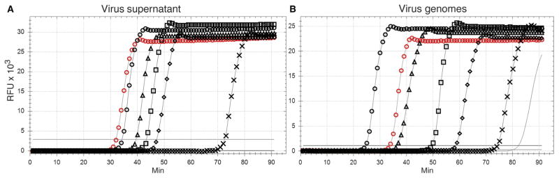
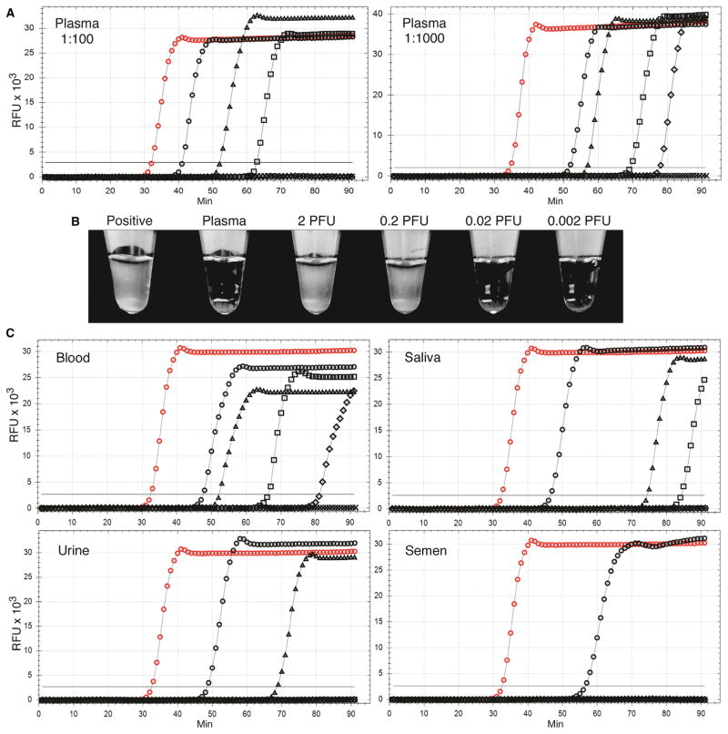
Understanding the dynamics of Zika virus transmission and formulating rational strategies for its control require precise diagnostic tools that are also appropriate for resource-poor environments. We have developed a rapid and sensitive loop-mediated isothermal amplification (LAMP) assay that distinguishes Zika viruses of Asian and African lineages. The assay does not detect chikungunya virus or flaviviruses such as dengue, yellow fever, or West Nile viruses. The assay conditions allowed direct detection of Zika virus RNA in cultured infected cells; in mosquitoes; in virus-spiked samples of human blood, plasma, saliva, urine, and semen; and in infected patient serum, plasma, and semen samples without the need for RNA isolation or reverse transcription. The assay offers rapid, specific, sensitive, and inexpensive detection of the Asian-lineage Zika virus strain that is currently circulating in the Western hemisphere, and can also detect the African-lineage Zika virus strain using separate, specific primers.
Copyright © 2017, American Association for the Advancement of Science.
Conflict of interest statement
Figures
Comment in
-
Switch-on the LAMP to spot Zika.Ann Transl Med. 2017 Dec;5(24):500. doi: 10.21037/atm.2017.10.19. Ann Transl Med. 2017. PMID: 29299461 Free PMC article. No abstract available.
-
LAMP assay for specific detection of Asian and African lineage Zika virus: will it meet the expectations?Ann Transl Med. 2018 Feb;6(3):53. doi: 10.21037/atm.2017.11.25. Ann Transl Med. 2018. PMID: 29610745 Free PMC article. No abstract available.
Similar articles
-
Point of sampling detection of Zika virus within a multiplexed kit capable of detecting dengue and chikungunya.BMC Infect Dis. 2017 Apr 20;17(1):293. doi: 10.1186/s12879-017-2382-0. BMC Infect Dis. 2017. PMID: 28427352 Free PMC article.
-
Development and evaluation of a visible reverse transcription-loop-mediated isothermal amplification (RT-LAMP) for the detection of Asian lineage ZIKV in field-caught mosquitoes.Acta Trop. 2022 Dec;236:106691. doi: 10.1016/j.actatropica.2022.106691. Epub 2022 Sep 11. Acta Trop. 2022. PMID: 36103950
-
Development of Universal and Lineage-Specific Primer Sets for Rapid Detection of the Zika Virus (ZIKV) in Blood and Urine Samples Using One-Step Reverse Transcription Loop-Mediated Isothermal Amplification (RT-LAMP).Jpn J Infect Dis. 2020 Mar 24;73(2):153-156. doi: 10.7883/yoken.JJID.2019.073. Epub 2019 Oct 31. Jpn J Infect Dis. 2020. PMID: 31666491
-
Flaviviruses and the Traveler: Around the World and to Your Stage. A Review of West Nile, Yellow Fever, Dengue, and Zika Viruses for the Practicing Pathologist.Mod Pathol. 2023 Jun;36(6):100188. doi: 10.1016/j.modpat.2023.100188. Epub 2023 Apr 13. Mod Pathol. 2023. PMID: 37059228 Review.
-
Loop-Mediated Isothermal Amplification (LAMP) for the Diagnosis of Zika Virus: A Review.Viruses. 2019 Dec 23;12(1):19. doi: 10.3390/v12010019. Viruses. 2019. PMID: 31877989 Free PMC article. Review.
Cited by
-
Laboratory diagnosis of SARS-CoV-2: available approaches and limitations.New Microbes New Infect. 2020 Jun 14;36:100713. doi: 10.1016/j.nmni.2020.100713. eCollection 2020 Jul. New Microbes New Infect. 2020. PMID: 32607246 Free PMC article. Review.
-
Single-cell nucleic acid profiling in droplets (SNAPD) enables high-throughput analysis of heterogeneous cell populations.Nucleic Acids Res. 2021 Oct 11;49(18):e103. doi: 10.1093/nar/gkab577. Nucleic Acids Res. 2021. PMID: 34233007 Free PMC article.
-
Genetics and genomics of SARS-CoV-2: A review of the literature with the special focus on genetic diversity and SARS-CoV-2 genome detection.Genomics. 2021 Jan;113(1 Pt 2):1221-1232. doi: 10.1016/j.ygeno.2020.09.059. Epub 2020 Sep 30. Genomics. 2021. PMID: 33007398 Free PMC article. Review.
-
Tracking virus outbreaks in the twenty-first century.Nat Microbiol. 2019 Jan;4(1):10-19. doi: 10.1038/s41564-018-0296-2. Epub 2018 Dec 13. Nat Microbiol. 2019. PMID: 30546099 Free PMC article. Review.
-
Rapid colorimetric detection of Zika virus from serum and urine specimens by reverse transcription loop-mediated isothermal amplification (RT-LAMP).PLoS One. 2017 Sep 25;12(9):e0185340. doi: 10.1371/journal.pone.0185340. eCollection 2017. PLoS One. 2017. PMID: 28945787 Free PMC article.
References
-
- Petersen LR, Jamieson DJ, Powers AM, Honein MA. Zika virus. N Engl J Med. 2016;374:1552–1563. - PubMed
Publication types
MeSH terms
Substances
Grants and funding
LinkOut - more resources
Full Text Sources
Other Literature Sources
