. 2017 May 4;2(9):e89996.
doi: 10.1172/jci.insight.89996.

p63+ ureteric bud tip cells are progenitors of intercalated cells

Affiliations

p63+ ureteric bud tip cells are progenitors of intercalated cells

Samir S El-Dahr et al. JCI Insight. .

Abstract

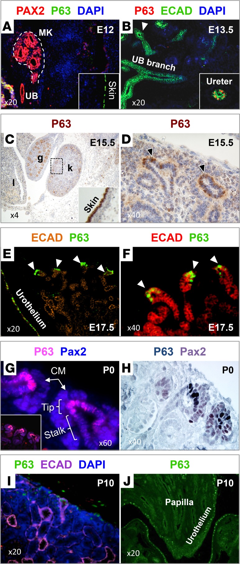
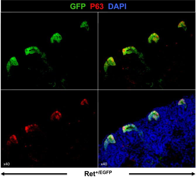
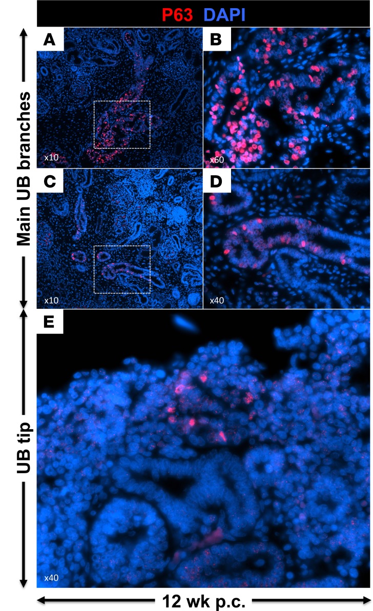
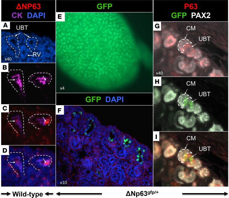
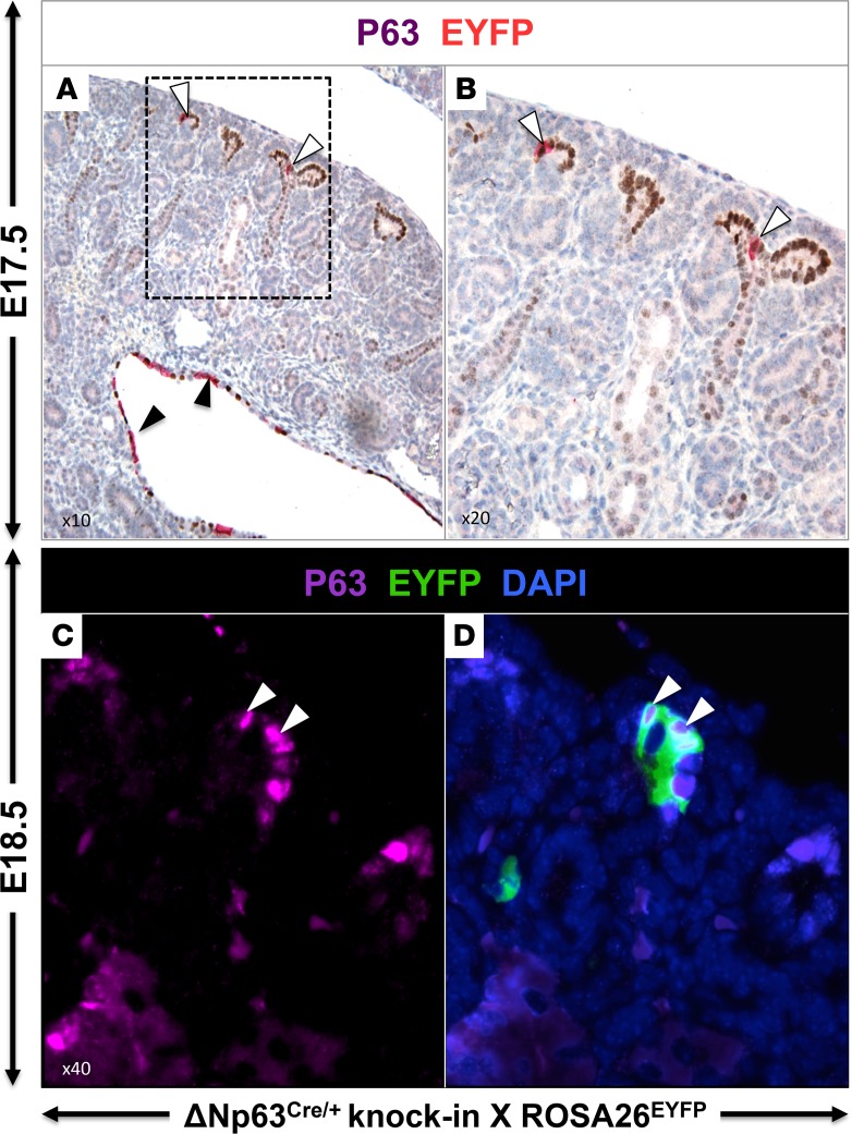
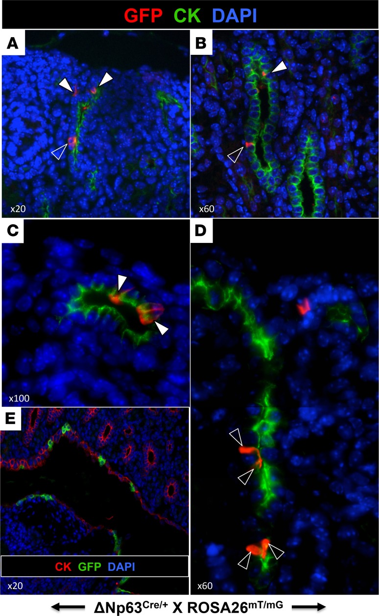
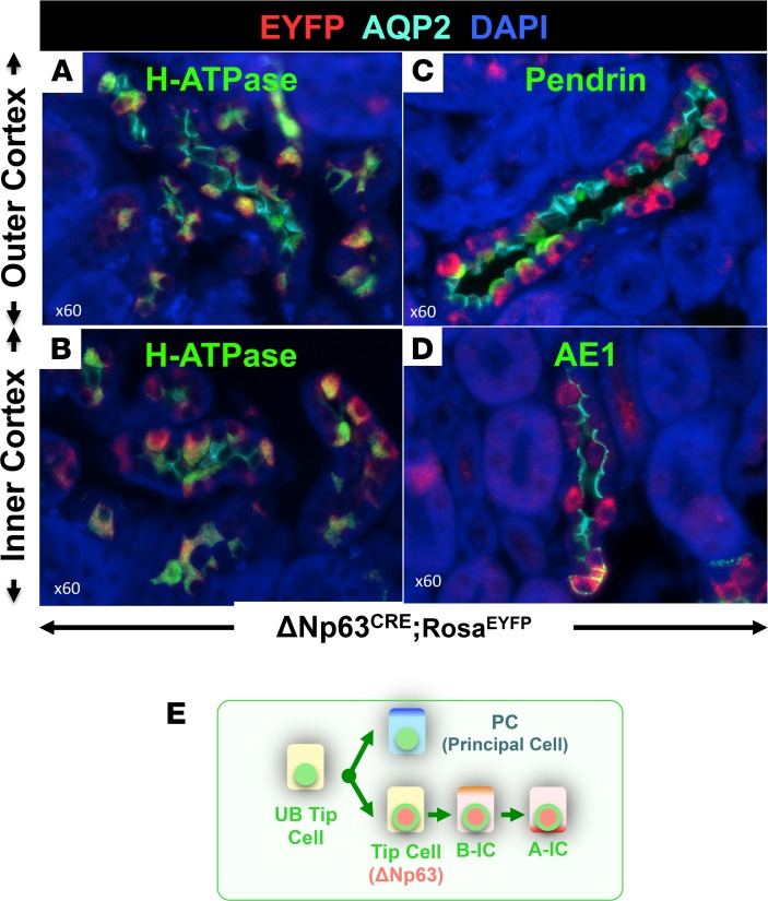
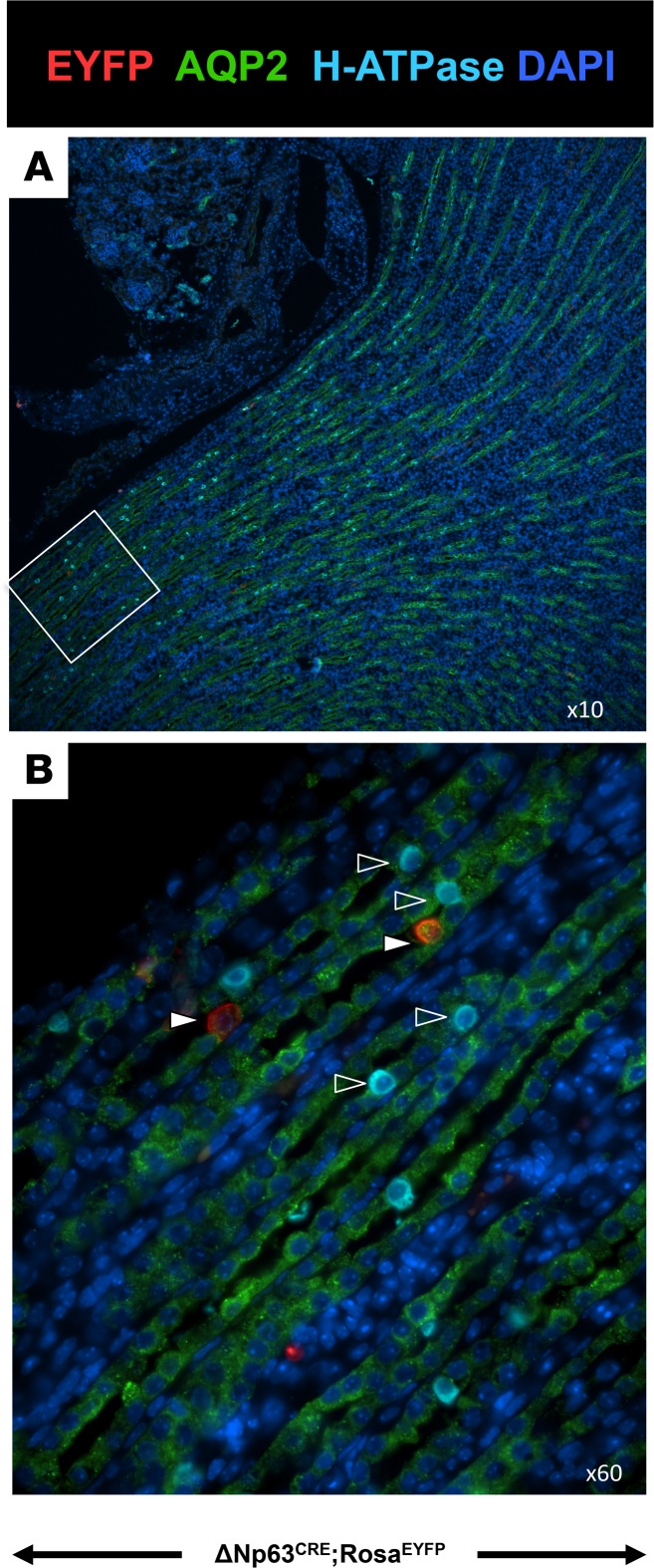
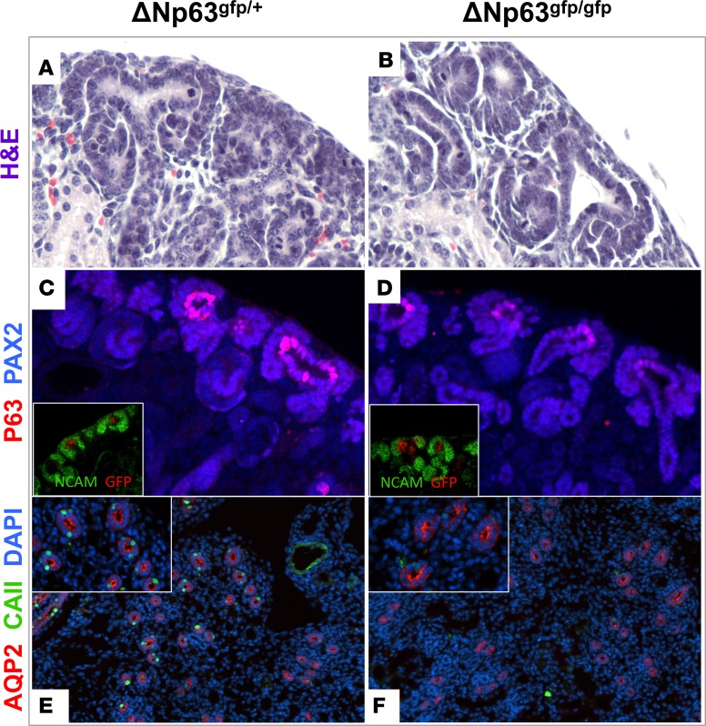
During renal branching morphogenesis, ureteric bud tip cells (UBTC) serve as the progenitor epithelium for all cell types of the collecting duct. While the transcriptional circuitry of ureteric bud (UB) branching has been intensively studied, the transcriptional control of UBTC differentiation has been difficult to ascertain. This is partly due to limited knowledge of UBTC-specific transcription factors that mark the progenitor state. Here, we identify the transcription factor p63 (also known as TP63), a master regulator of basal stem cells in stratified epithelia, as a specific marker of mouse and human UBTC. Nuclear p63 marks Ret+ UBTC transiently and is silenced by the end of nephrogenesis. Lineage tracing revealed that a subset of UBTC expressing the ΔNp63 isoform (N-terminus truncated p63) is dedicated to generating cortical intercalated cells. Germline targeting of ΔNp63 in mice caused a marked reduction in intercalated cells near the time of birth, indicating that p63 not only marks UBTC, but also is essential for their differentiation. We conclude that the choice of UBTC progenitors to differentiate is determined earlier than previously recognized and that UBTC progenitors are prepatterned and fate restricted. These findings prompt the rethinking of current paradigms of collecting duct differentiation and may have implications for regenerative renal medicine.

Keywords: Development; Nephrology.


Conflict of interest statement

Conflict of interest: The authors have declared that no conflict of interest exists.

Figures

Figure 1
Figure 1. p63 marks ureteric bud tip progenitors.
Immunofluorescence (A, B, EG, I, and J) and immunohistochemistry (C, D, and H) using anti-p63 antibody (4A4). (A) At E12, p63 (green) is expressed in skin (inset) but not in the ureteric bud (UB) or UB branches. Pax2 (red) is used as a marker of the nephric lineage. MK, metanephric kidney. (B) At E13.5, p63 (red) is expressed in the UB epithelium but not its branches or tips (arrowhead). E-cadherin (ECAD, green) is used as marker of the UB epithelium. (C and D) E15.5 marks the onset of p63 expression in the UB tip cells (arrowheads). L, liver; g, gonad; k, kidney. (E and F) On E17.5, UB tip–restricted expression of p63 is clearly evident. Arrowheads denote p63+ UB tip cells. (G and H) At P0, p63 marks UB tip cells but not the cap mesenchyme (CM) or UB stalk. (I and J) p63 is silenced after P5. At P10, p63 is not detectable in the maturing collecting ducts but remains present in the pelvic urothelium (n = 3–5 animals per age group). Original magnification, ×4 (C); ×20 (A, B, E, I, and J); ×40 (D, F, H, and all insets); ×60 (G).
Figure 2
Figure 2. p63 labels a subset of Ret+ ureteric bud tip cells.
Section immunofluorescence in P1 kidneys of Ret+/EGFP mice. Ret/EGFP (green) marks the entire ureteric bud (UB) tip domain, whereas p63 marks a subset of EGFP+ cells. The distribution of p63+ cells in the UB tip is not associated with branching points (n = 3). Original magnification, ×40.
Figure 3
Figure 3. p63 is expressed in ureteric bud tip cells of human fetal kidneys.
A tissue section from a 12-week-old human fetus stained with p63 (4A4) antibody. (A and B) Low- and high-power images of the central ureteric bud (UB) branches, which express p63. (CE) p63 is expressed in cortical collecting ducts and in the tip domain (n = 1 fetal kidney). Original magnification, ×10 (A and C); ×40 (D and E); ×60 (B).
Figure 4
Figure 4. ΔNp63 is expressed in a subset of ureteric bud tip cells.
(AD) Section immunofluorescence of an E17.5 kidney using ΔNp63-specific antibody. Staining for cytokeratin (CK) identifies the ureteric bud (UB) branches. Arrows point to ΔNp63+ cells in BD. RV, renal vesicle. (E) Whole mount GFP fluorescence in an E18.5 kidney from a ΔNp63+/gfp knockin mouse. Note the tip expression of GFP. (F) Section immunofluorescence using anti-GFP antibody in an E18.5 ΔNp63+/gfp kidney showing UB tip–specific expression of GFP. (GI) Section immunofluorescence using anti-GFP, p63, and Pax2 antibodies in an E18.5 ΔNp63+/gfp kidney. p63 (red) labels the UB tip domain (UBT). Pax2 (gray) labels the UB tip (UBT) and surrounding cap mesenchyme (CM). GFP (green), representing ΔNp63, is expressed in a subset of p63+ ureteric bud tip cells (n = 2–5 animals/experiment). Original magnification, ×4 (E); ×10 (F); ×40 (AD and GI).
Figure 5
Figure 5. Fate tracing of UBTCΔNp63 in E17.5 ΔNp63+/Cre;ROSA26EYFP mice.
(A and B) Section immunohistochemistry. p63 (brown) strongly labels the ureteric bud tip cells (UBTC) and pelvic urothelium. EYFP (purple) labels 1–2 UBTC (white arrowheads) and patches of pelvic epithelial cells (black arrowheads). (C and D) Colocalization of p63 protein (purple) with EYFP (green) in UBTC (n = 5). Arrowheads point to p63+/EYFP+ cells. Original magnification, ×10 (A); ×20 (B); ×40 (C and D).
Figure 6
Figure 6. Fate tracing of UBTCΔNp63 in P1 ΔNp63+/Cre;ROSA26mT/mG mice.
Section immunofluorescence staining of the ureteric bud/collecting duct marker with cytokeratin (CK, green) and membrane-tethered GFP (red). Progeny of GFP+ UBTCΔNp63 pattern the cortical collecting ducts and are found in both tips (A and C; white arrowheads) and stalks (B and D, outlined arrowheads). (E) Low-power image of the deep medullary region showing the presence of GFP+ UBTCΔNp63 in the urothelium but not in adjacent medullary/papillary collecting ducts (n = 4). Original magnification, ×20 (A and E); ×60 (B and D); ×100 (C).
Figure 7
Figure 7. UBTCΔNp63 are precursors of cortical intercalated cells in the adult kidney.
(AD) ΔNp63+/Cre knockin mice were crossed to ROSA26EYFP reporter mice. Kidney sections from adult ΔNp63+/Cre;ROSA26EYFP mice were immunostained to label ΔNp63 daughter cells (EYFP), PC (AQP2), IC (vH-ATPase B1), A-type IC (AE1), and non–A-type IC (pendrin). EYFP+ cells stain positive for IC but not PC markers. (E) Model for ureteric bud tip cell (UBTC) differentiation: UBTC differentiate into ureteric bud (UB) precursors of PC and IC; ΔNp63+ cells located in UB tips differentiate to type B-IC, which in turn differentiate into type-A IC (n = 4). Original magnification, ×60.
Figure 8
Figure 8. Occasional EYFP+ UBTCΔNp63lineage are seen in the medulla of adult kidney.
However, these cells (red, white arrowheads) do not overlap with cells expressing the IC marker H-ATPase (blue, outlined arrowheads) (n = 4). Original magnification, ×10 (A); ×60 (B).
Figure 9
Figure 9. Homozygous deletion of ΔNp63 compromises IC differentiation.
(A and B) H&E staining showing intact overall structure of the nephrogenic zone in knockin ΔNp63gfp/gfp mice. (C and D) Double immunostaining with p63 (red) and Pax2 (purple) showing that loss of ΔNp63 diminishes expression of total p63 in UB tips. The insets show that NCAM (green) marks the induced cap mesenchyme and GFP labels ΔNp63 gene-targeted ureteric bud tip cells (UBTC). Loss of ΔNp63 has no effect on integrity of UBTC. (E and F) Double immunostaining of AQP2 (red) and carbonic anhydrase II (CAII, green) showing that targeted deletion of ΔNp63 results in loss of IC marker expression (n = 3 litters). Original magnification, ×20; ×40 (all insets).

Similar articles

Cited by

References

    1. Costantini F, Kopan R. Patterning a complex organ: branching morphogenesis and nephron segmentation in kidney development. Dev Cell. 2010;18(5):698–712. doi: 10.1016/j.devcel.2010.04.008. - DOI - PMC - PubMed
    1. Al-Awqati Q. Cell biology of the intercalated cell in the kidney. FEBS Lett. 2013;587(13):1911–1914. doi: 10.1016/j.febslet.2013.05.007. - DOI - PubMed
    1. Davis TK, Hoshi M, Jain S. To bud or not to bud: the RET perspective in CAKUT. Pediatr Nephrol. 2014;29(4):597–608. doi: 10.1007/s00467-013-2606-5. - DOI - PMC - PubMed
    1. Costantini F. Genetic controls and cellular behaviors in branching morphogenesis of the renal collecting system. Wiley Interdiscip Rev Dev Biol. 2012;1(5):693–713. doi: 10.1002/wdev.52. - DOI - PMC - PubMed
    1. Packard A, et al. Luminal mitosis drives epithelial cell dispersal within the branching ureteric bud. Dev Cell. 2013;27(3):319–330. doi: 10.1016/j.devcel.2013.09.001. - DOI - PMC - PubMed