

Long-term Levodopa Treatment Accelerates the Circadian Rhythm Dysfunction in a 6-hydroxydopamine Rat Model of Parkinson's Disease
- PMID: 28469105
- PMCID: PMC5421180
- DOI: 10.4103/0366-6999.204920
Long-term Levodopa Treatment Accelerates the Circadian Rhythm Dysfunction in a 6-hydroxydopamine Rat Model of Parkinson's Disease
Abstract
Background: Parkinson's disease (PD) patients with long-term levodopa (L-DOPA) treatment are suffering from severe circadian dysfunction. However, it is hard to distinguish that the circadian disturbance in patients is due to the disease progression itself, or is affected by L-DOPA replacement therapy. This study was to investigate the role of L-DOPA on the circadian dysfunction in a rat model of PD.
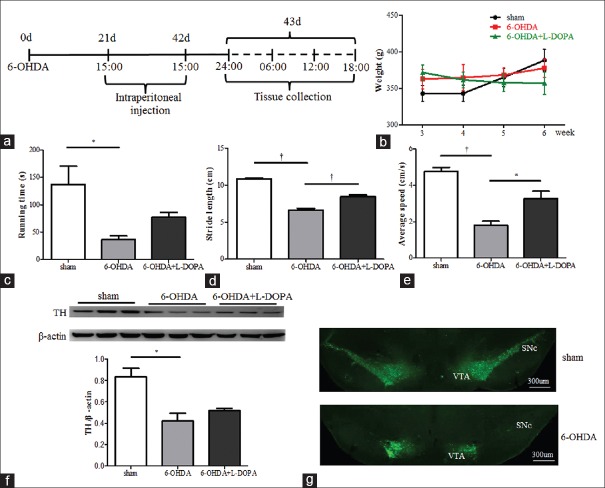
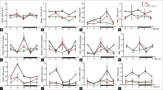
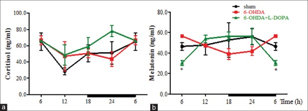
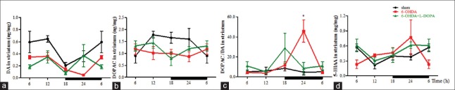
Methods: The rat model of PD was constructed by a bilateral striatal injection with 6-hydroxydopamine (6-OHDA), followed by administration of saline or 25 mg/kg L-DOPA for 21 consecutive days. Rotarod test, footprint test, and open-field test were carried out to evaluate the motor function. Striatum, suprachiasmatic nucleus (SCN), liver, and plasma were collected at 6:00, 12:00, 18:00, and 24:00. Quantitative real-time polymerase chain reaction was used to examine the expression of clock genes. Enzyme-linked immunosorbent assay was used to determine the secretion level of cortisol and melatonin. High-performance liquid chromatography was used to measure the neurotransmitters. Analysis of variance was used for data analysis.
Results: L-DOPA alleviated the motor deficits induced by 6-OHDA lesions in the footprint and open-field test ( P < 0.01, P < 0.001, respectively). After L-DOPA treatment, Bmal1 decreased in the SCN compared with 6-OHDA group at 12:00 ( P < 0.01) and 24:00 ( P < 0.001). In the striatum, the expression of Bmal1, Rorα was lower than that in the 6-OHDA group at 18:00 (P < 0.05) and L-DOPA seemed to delay the peak of Per2 to 24:00. In liver, L-DOPA did not affect the rhythmicity and expression of these clock genes (P > 0.05). In addition, the cortisol secretion was increased (P > 0.05), but melatonin was further inhibited after L-DOPA treatment at 6:00 (P < 0.01).
Conclusions: In the circadian system of advanced PD rat models, circadian dysfunction is not only contributed by the degeneration of the disease itself but also long-term L-DOPA therapy may further aggravate it.
Conflict of interest statement
There are no conflicts of interest.
Figures
Similar articles
-
Effects of L-Dopa on circadian rhythms of 6-OHDA striatal lesioned rats: a radiotelemetric study.Chronobiol Int. 2010 Jan;27(2):251-64. doi: 10.3109/07420521003664213. Chronobiol Int. 2010. PMID: 20370468
-
Levodopa/benserazide microsphere (LBM) prevents L-dopa induced dyskinesia by inactivation of the DR1/PKA/P-tau pathway in 6-OHDA-lesioned Parkinson's rats.Sci Rep. 2014 Dec 16;4:7506. doi: 10.1038/srep07506. Sci Rep. 2014. PMID: 25511986 Free PMC article.
-
L-3,4-Dihydroxyphenylalanine Recovers Circadian Rhythm Disturbances in the Rat Models of Parkinson's Disease by Regulating the D1R-ERK1/2-mTOR Pathway.Front Aging Neurosci. 2021 Aug 19;13:719885. doi: 10.3389/fnagi.2021.719885. eCollection 2021. Front Aging Neurosci. 2021. PMID: 34489685 Free PMC article.
-
Intranigral administration of substance P receptor antagonist attenuated levodopa-induced dyskinesia in a rat model of Parkinson's disease.Exp Neurol. 2015 Sep;271:168-74. doi: 10.1016/j.expneurol.2015.05.007. Epub 2015 May 20. Exp Neurol. 2015. PMID: 26001615
-
Electroanalytical Overview: The Determination of Levodopa (L-DOPA).ACS Meas Sci Au. 2023 Feb 3;3(2):84-97. doi: 10.1021/acsmeasuresciau.2c00071. eCollection 2023 Apr 19. ACS Meas Sci Au. 2023. PMID: 37090256 Free PMC article. Review.
Cited by
-
Epigenetic events influencing the biological clock: Panacea for neurodegeneration.Heliyon. 2024 Oct 2;10(19):e38836. doi: 10.1016/j.heliyon.2024.e38836. eCollection 2024 Oct 15. Heliyon. 2024. PMID: 39430507 Free PMC article. Review.
-
Novel Treatment Strategies for the Nervous System: Circadian Clock Genes, Non-coding RNAs, and Forkhead Transcription Factors.Curr Neurovasc Res. 2018;15(1):81-91. doi: 10.2174/1567202615666180319151244. Curr Neurovasc Res. 2018. PMID: 29557749 Free PMC article. Review.
-
The Role of Circadian Rhythm in Neurological Diseases: A Translational Perspective.Aging Dis. 2024 Aug 1;15(4):1565-1587. doi: 10.14336/AD.2023.0921. Aging Dis. 2024. PMID: 37815902 Free PMC article. Review.
-
Cognitive impairment with diabetes mellitus and metabolic disease: innovative insights with the mechanistic target of rapamycin and circadian clock gene pathways.Expert Rev Clin Pharmacol. 2020 Jan;13(1):23-34. doi: 10.1080/17512433.2020.1698288. Epub 2020 Jan 3. Expert Rev Clin Pharmacol. 2020. PMID: 31794280 Free PMC article. Review.
-
Cortisol levels, motor, cognitive and behavioral symptoms in Parkinson's disease: a systematic review.J Neural Transm (Vienna). 2019 Mar;126(3):219-232. doi: 10.1007/s00702-018-1947-4. Epub 2018 Oct 29. J Neural Transm (Vienna). 2019. PMID: 30374595
References
-
- Zhang ZX, Roman GC, Hong Z, Wu CB, Qu QM, Huang JB, et al. Parkinson's disease in China: Prevalence in Beijing, Xian, and Shanghai. Lancet. 2005;365:595–7. doi: 10.1016/S0140-6736(05)17909-4. - PubMed
-
- Bonuccelli U, Del Dotto P, Lucetti C, Petrozzi L, Bernardini S, Gambaccini G, et al. Diurnal motor variations to repeated doses of levodopa in Parkinson's disease. Clin Neuropharmacol. 2000;23:28–33. - PubMed
-
- Faludi B, Janszky J, Komoly S, Kovács N. Sleep disturbances in Parkinson's disease: Characteristics, evaluation and therapeutic approaches. Orv Hetil. 2015;156:1091–9. doi: 10.1556/650.2015.30191. - PubMed
-
- Struck LK, Rodnitzky RL, Dobson JK. Circadian fluctuations of contrast sensitivity in Parkinson's disease. Neurology. 1990;40(3 Pt 1):467–70. - PubMed
MeSH terms
Substances
LinkOut - more resources
Full Text Sources
Other Literature Sources
