

Subventricular zone involvement in Glioblastoma - A proteomic evaluation and clinicoradiological correlation
- PMID: 28469129
- PMCID: PMC5431125
- DOI: 10.1038/s41598-017-01202-8
Subventricular zone involvement in Glioblastoma - A proteomic evaluation and clinicoradiological correlation
Abstract
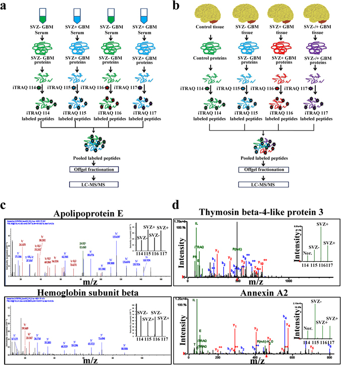
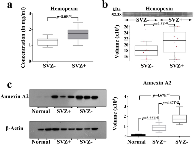
Glioblastoma multiforme (GBM), the most malignant of all gliomas is characterized by a high degree of heterogeneity and poor response to treatment. The sub-ventricular zone (SVZ) is the major site of neurogenesis in the brain and is rich in neural stem cells. Based on the proximity of the GBM tumors to the SVZ, the tumors can be further classified into SVZ+ and SVZ-. The tumors located in close contact with the SVZ are classified as SVZ+, while the tumors located distantly from the SVZ are classified as SVZ-. To gain an insight into the increased aggressiveness of SVZ+ over SVZ- tumors, we have used proteomics techniques like 2D-DIGE and LC-MS/MS to investigate any possible proteomic differences between the two subtypes. Serum proteomic analysis revealed significant alterations of various acute phase proteins and lipid carrying proteins, while tissue proteomic analysis revealed significant alterations in cytoskeletal, lipid binding, chaperone and cell cycle regulating proteins, which are already known to be associated with disease pathobiology. These findings provide cues to molecular basis behind increased aggressiveness of SVZ+ GBM tumors over SVZ- GBM tumors and plausible therapeutic targets to improve treatment modalities for these highly invasive tumors.
Conflict of interest statement
The authors declare that they have no competing interests.
Figures


Similar articles
-
Distant origin of glioblastoma recurrence: neural stem cells in the subventricular zone serve as a source of tumor reconstruction after primary resection.Mol Cancer. 2025 Mar 4;24(1):64. doi: 10.1186/s12943-025-02273-2. Mol Cancer. 2025. PMID: 40033380 Free PMC article.
-
Glioblastomas located in proximity to the subventricular zone (SVZ) exhibited enrichment of gene expression profiles associated with the cancer stem cell state.J Neurooncol. 2020 Jul;148(3):455-462. doi: 10.1007/s11060-020-03550-4. Epub 2020 Jun 15. J Neurooncol. 2020. PMID: 32556864
-
Comparative analyses identify molecular signature of MRI-classified SVZ-associated glioblastoma.Cell Cycle. 2017 Apr 18;16(8):765-775. doi: 10.1080/15384101.2017.1295186. Epub 2017 Feb 22. Cell Cycle. 2017. PMID: 28278055 Free PMC article.
-
Overlapping migratory mechanisms between neural progenitor cells and brain tumor stem cells.Cell Mol Life Sci. 2019 Sep;76(18):3553-3570. doi: 10.1007/s00018-019-03149-7. Epub 2019 May 17. Cell Mol Life Sci. 2019. PMID: 31101934 Free PMC article. Review.
-
Influence of glioblastoma contact with the lateral ventricle on survival: a meta-analysis.J Neurooncol. 2017 Jan;131(1):125-133. doi: 10.1007/s11060-016-2278-7. Epub 2016 Sep 19. J Neurooncol. 2017. PMID: 27644688 Free PMC article. Review.
Cited by
-
RND1 regulates migration of human glioblastoma stem-like cells according to their anatomical localization and defines a prognostic signature in glioblastoma.Oncotarget. 2018 Sep 18;9(73):33788-33803. doi: 10.18632/oncotarget.26082. eCollection 2018 Sep 18. Oncotarget. 2018. PMID: 30333910 Free PMC article.
-
AN1-type zinc finger protein 3 (ZFAND3) is a transcriptional regulator that drives Glioblastoma invasion.Nat Commun. 2020 Dec 11;11(1):6366. doi: 10.1038/s41467-020-20029-y. Nat Commun. 2020. PMID: 33311477 Free PMC article.
-
High dose vs low dose irradiation of the subventricular zone in patients with glioblastoma-a systematic review and meta-analysis.Cancer Manag Res. 2019 Jul 18;11:6741-6753. doi: 10.2147/CMAR.S206033. eCollection 2019. Cancer Manag Res. 2019. PMID: 31410064 Free PMC article.
-
The influence of subventricular zone involvement in extent of resection and tumor growth pattern of glioblastoma.Innov Surg Sci. 2020 Aug 27;5(3-4):127-132. doi: 10.1515/iss-2020-0011. eCollection 2020 Sep. Innov Surg Sci. 2020. PMID: 34966832 Free PMC article.
-
Role and mechanism of neural stem cells of the subventricular zone in glioblastoma.World J Stem Cells. 2021 Jul 26;13(7):877-893. doi: 10.4252/wjsc.v13.i7.877. World J Stem Cells. 2021. PMID: 34367482 Free PMC article. Review.
References
Publication types
MeSH terms
Substances
LinkOut - more resources
Full Text Sources
Other Literature Sources
Medical
