. 2017 May 3;7(1):1431.
doi: 10.1038/s41598-017-01486-w.

Revisiting antithrombotic therapeutics; sculptin, a novel specific, competitive, reversible, scissile and tight binding inhibitor of thrombin

Affiliations

Revisiting antithrombotic therapeutics; sculptin, a novel specific, competitive, reversible, scissile and tight binding inhibitor of thrombin

Asif Iqbal et al. Sci Rep. .

Abstract

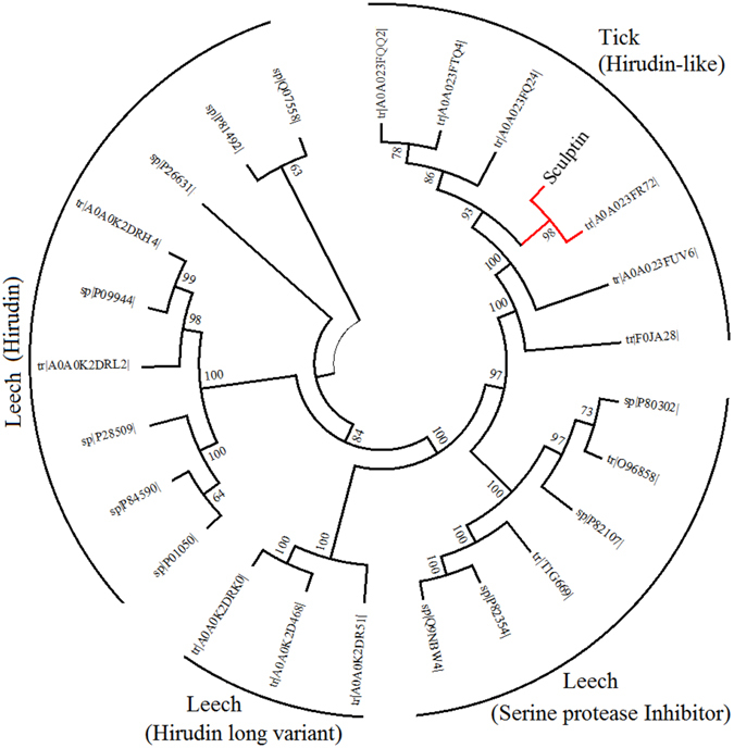
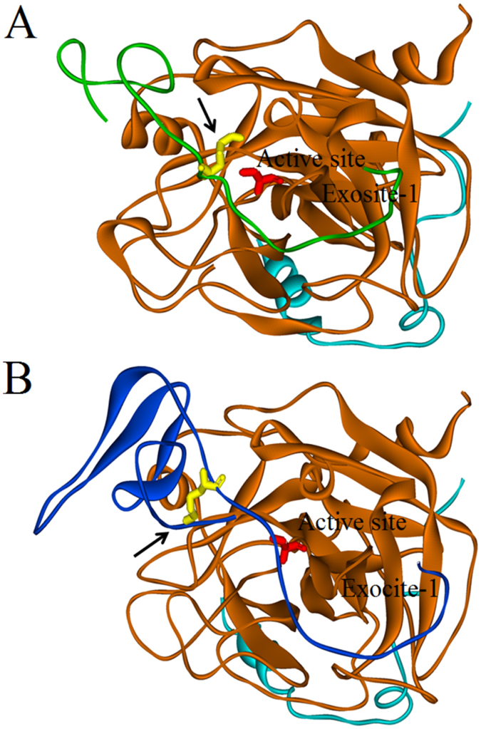
Thrombin is a multifunctional enzyme with a key role in the coagulation cascade. Its functional modulation can culminate into normal blood coagulation or thrombosis. Thus, the identification of novel potent inhibitors of thrombin are of immense importance. Sculptin is the first specific thrombin inhibitor identified in the transcriptomics analysis of tick's salivary glands. It consists of 168 residues having four similar repeats and evolutionary diverged from hirudin. Sculptin is a competitive, specific and reversible inhibitor of thrombin with a Ki of 18.3 ± 1.9 pM (k on 4.04 ± 0.03 × 10⁷ M-1 s-1 and k off 0.65 ± 0.04 × 10⁻³ s-1). It is slowly consumed by thrombin eventually losing its activity. Contrary, sculptin is hydrolyzed by factor Xa and each polypeptide fragment is able to inhibit thrombin independently. A single domain of sculptin alone retains ~45% of inhibitory activity, which could bind thrombin in a bivalent fashion. The formation of a small turn/helical-like structure by active site binding residues of sculptin might have made it a more potent thrombin inhibitor. In addition, sculptin prolongs global coagulation parameters. In conclusion, sculptin and its independent domain(s) have strong potential to become novel antithrombotic therapeutics.


Conflict of interest statement

The author(s) see no competing financial interests. However, this molecule is submitted for patenting with INPI number BR 102017005783-6.

Figures

Figure 1
Figure 1
Phylogenetic analysis of Sculptin. Protein sequences of thrombin inhibitors from ticks and leeches were retrieved from Swiss-Prot/TrEMBL database (www.uniprot.org) and the phylogenetic profile was inferred using the Neighbor-Joining method using MEGA 7.0. The bootstrap consensus were 100 and cut-off value for condensed tree was 60% collapsed bootstrap replicates (see, experimental procedures). The accession number of each sequence is given and query position is highlighted in red. A single domain of sculptin was taken in to account during phylogeny construction.
Figure 2
Figure 2
Sculptin specificity for thrombin and its dose-dependency and IC50 value for thrombin inhibition. (A) The inhibition of serine proteases by sculptin. Serine protease (100 pM; thrombin, plasmin, trypsin or factor Xa) was incubated with sculptin (1, 100 and 200 nM) in 50 mM phosphate buffer containing 150 mM NaCl and 0.1% PEG 6000, pH 7.4 for 6 h at 37 °C. After addition of the corresponding chromogenic substrate to the reaction mixture, its hydrolysis was monitored at 405 nm. For factor Xa activity, the buffer contained 50 µM phosphatidylserine and phosphatidylcholine. The inset in (A) shows the SDSPAGE of purified recombinant sculptin, which was used in the experiments. (B) Typical curves for hydrolysis of S-2238 chromogenic substrate (15 µM) by 0.1 nM thrombin in the absence (trace a) or presence of sculptin (trace b, 15 pM; trace c, 30 pM; trace d, 60 pM and trace e, 100 pM) in 50 mM phosphate buffer containing 150 mM NaCl and 0.1% PEG 6000, pH 7.4 at 37 °C. (C) Residual activity of thrombin in presence of increasing concentration of sculptin. (D) Dose-response curve for thrombin inhibition by sculptin. The percentage of thrombin inhibition was plotted versus the log of sculptin concentration. The experimental condition of (C) and (D) is the same as in (B). The results shown in (C) and (D) correspond to the mean ± standard deviation values acquired in three independent experiments.
Figure 3
Figure 3
Kinetics of inhibition of thrombin by sculptin. (A) Typical progress curves for hydrolysis of S-2238 chromogenic substrate by 0.1 nM thrombin in absence (trace a) and presence of sculptin (trace b, 20 pM; trace c, 40 pM; trace d, 60 pM and trace e, 80 pM) in 50 mM phosphate buffer containing 150 mM NaCl and 0.1% PEG 6000, pH 7.4 at 37 °C. Reactions were started with the addition of thrombin to the mixture containing sculptin and S-2238. (B) The Hanes-Woolf plot for the inhibition of thrombin by sculptin (trace a, 0 pM; trace b, 20 pM; trace c, 40 pM and trace d, 80 pM). The substrate concentrations divided by its corresponding initial velocities of thrombin inhibition by sculptin were plotted vs sculptin concentration using Eq. (2). (C) The intercept of the (B) was plotted versus respective concentration to obtain Ki. (D) The nonlinear regression for competitive inhibition using Eqs (3) and (4). The initial velocity of thrombin inhibition in absence (trace a) and presence of sculptin (trace b, 20 pM; trace c, 40 pM; trace d, 60 pM and trace e, 80 pM) at different substrate concentrations. The experimental condition of (B,C) and (D) is the same as (A). The data in (B,C) and (D) correspond to the mean ± standard deviation values acquired in five independent experiments.
Figure 4
Figure 4
Relationship between the apparent first-order rate and the concentration of tight binding inhibitor sculptin. (A) Typical progress curves for hydrolysis of 15 µM S-2238 chromogenic substrate by 0.1 nM thrombin in absence (trace a) and presence of sculptin (trace b, 10 pM; trace c, 30 pM; trace d, 70 pM; trace e, 100 pM; trace f, 200 pM and trace g, 500 pM) in 50 mM phosphate buffer containing 150 mM NaCl and 0.1% PEG 6000, pH 7.4 at 37 °C. Reactions were started with addition of thrombin to the mixture containing sculptin and S-2238. (B) Steady-state velocity of thrombin with respect to sculptin concentration. The inset shows the determination of the apparent dissociation constant, Ki*, from steady state velocities. The data was fitted into a linear regression to obtain Ki. (C) Calculation of the dissociation constant from kobs. Progress curves were produced with 15 µM S-2238, 0–50 pM sculptin, and 100 pM thrombin. The apparent first-order rate constant was calculated using a nonlinear regression fit, where the intercept and slope is kon and koff respectively. The experimental condition of (B) and (C) is the same as (A). The data in (B) and (C) correspond to the mean ± standard deviation values acquired in five independent experiments.
Figure 5
Figure 5
Degradation of sculptin by serine proteases and its thrombin inhibition activity. Sculptin (10 µM) was incubated without or with 1 µM of serine protease (thrombin, plasmin, trypsin or factor Xa) in 50 mM phosphate buffer containing 150 mM NaCl and 0.1% PEG 6000, pH 7.4 for 4, 6, 7 or 18 h at 37 °C. The reaction mixtures (20 µl) were separated by SDS-PAGE. (A) SDS-PAGE (15%) of sculptin hydrolysis by serine proteases after 6 h incubation. (B) Percent thrombin inhibition by sculptin after 6 h incubation with serine proteases (see experimental procedures). (C) SDS-PAGE (15%) of sculptin hydrolysis by serine proteases after 18 h incubation. (D) Percent thrombin inhibition by sculptin after 18 h incubation with serine protease (see experimental procedures). The numbering of (B) and (D) correspond to the numbering of (A) and (C) respectively and sculptin control is represented by CTRL. Sculptin (lane 1); thrombin (lane 2) and thrombin with sculptin (lane 3); plasmin (lane 4) and plasmin with sculptin (lane 5); trypsin (lane 6) and trypsin with sculptin (lane 7); factor Xa (lane 8) and factor Xa with sculptin (lane 9) and protein marker (lane 10; in A). (E) Identification of thrombin cleavage sites in sculptin after 7 h of incubation. (F) Identification of factor Xa cleavage sites in sculptin after 4 h of incubation. Thrombin and Factor Xa cleavage sites in sculptin sequence are given in Figs S1 and S2 respectively. The experimental procedure for (CE) and (F) was same for (A) and (B) except incubation time and type of serine protease used. The results shown in (B) and (D) correspond to the mean ± standard deviation values acquired in three independent experiments; ns nonsignificant, ***p < 0.001 and *p < 0.05.
Figure 6
Figure 6
Thrombin inhibition activity of sculptin fragments generated by factor Xa. Sculptin (10 µM) was incubated without or with 1 µM of factor Xa in 50 mM phosphate buffer containing 150 mM NaCl, and 50 µM phosphatidylserine and phosphatidylcholine, pH 7.4 for 6 h at 37 °C. (A) The reaction mixtures were separated by reversed phase C-18 HPLC column. (B) The collected peaks (H1–H5) were subjected to MALDI-TOF MS and thrombin inhibition assay (see Table 1, for sequence of the corresponding peptides). (C) Typical progress curves for hydrolysis of 15 µM S-2238 chromogenic substrate by 0.1 nM thrombin in the absence (trace Ctrl) and presence of 100 pM of sculptin fragment (traces H1 and H3) or intact sculptin (trance Scpt) in 50 mM phosphate buffer containing 150 mM NaCl and 0.1% PEG 6000, pH 7.4 at 37 °C. (D) Percent thrombin inhibition by sculptin and its fragments. The reaction conditions of (D) are same as in (C). The results shown in (D) correspond to the mean ± standard deviation values acquired in three independent experiments; **p < 0.01 and *p < 0.05.
Figure 7
Figure 7
Cartoon (Solid ribbon) representation of comparative structure of sculptin (single domain) and crystal structure of hirudin bound to thrombin. The brown and cyan color represent the heavy and light chains of thrombin respectively. (A) Sculptin in green bound to thrombin. (B) Hirudin in blue bound to thrombin. Lys residue of the inhibitors is shown in yellow and Ser195 residue of the thrombin active site is presented in red. The arrow indicates the active site binding pocket of the thrombin occupied by Lys23 residue of sculptin (B) and by the N-terminal region of hirudin (A).

References

    1. Posma JJN, Posthuma JJ, Spronk HMH. Coagulation and non-coagulation effects of thrombin. J. Thromb. Haemost. 2016;14:1908–1916. doi: 10.1111/jth.13441. - DOI - PubMed
    1. Lee CJ, Ansell JE. Direct thrombin inhibitors: Parenteral and oral inhibitors of thrombin activity. Br. J. Clin. Pharmacol. 2011;72:581–592. doi: 10.1111/j.1365-2125.2011.03916.x. - DOI - PMC - PubMed
    1. Crawley JTB, Zanardelli S, Chion CKNK, Lane DA. The central role of thrombin in hemostasis: Central role of thrombin in hemostasis. J. Thromb. Haemost. 2007;5:95–101. doi: 10.1111/j.1538-7836.2007.02500.x. - DOI - PubMed
    1. Siller-Matula JM, Schwameis M, Blann A, Mannhalter C, Jilma B. Thrombin as a multi-functional enzyme: Focus on in vitro and in vivo effects. Thromb. Haemost. 2011;106:1020–1033. doi: 10.1160/TH10-11-0711. - DOI - PubMed
    1. Coppens M, Eikelboom JW, Gustafsson D, Weitz JI, Hirsh J. Translational Success Stories: Development of Direct Thrombin Inhibitors. Circ. Res. 2012;111:920–929. doi: 10.1161/CIRCRESAHA.112.264903. - DOI - PubMed

Publication types

MeSH terms