

Protective effect of antioxidants on the pre-maturation aging of mouse oocytes
- PMID: 28469172
- PMCID: PMC5431116
- DOI: 10.1038/s41598-017-01609-3
Protective effect of antioxidants on the pre-maturation aging of mouse oocytes
Abstract
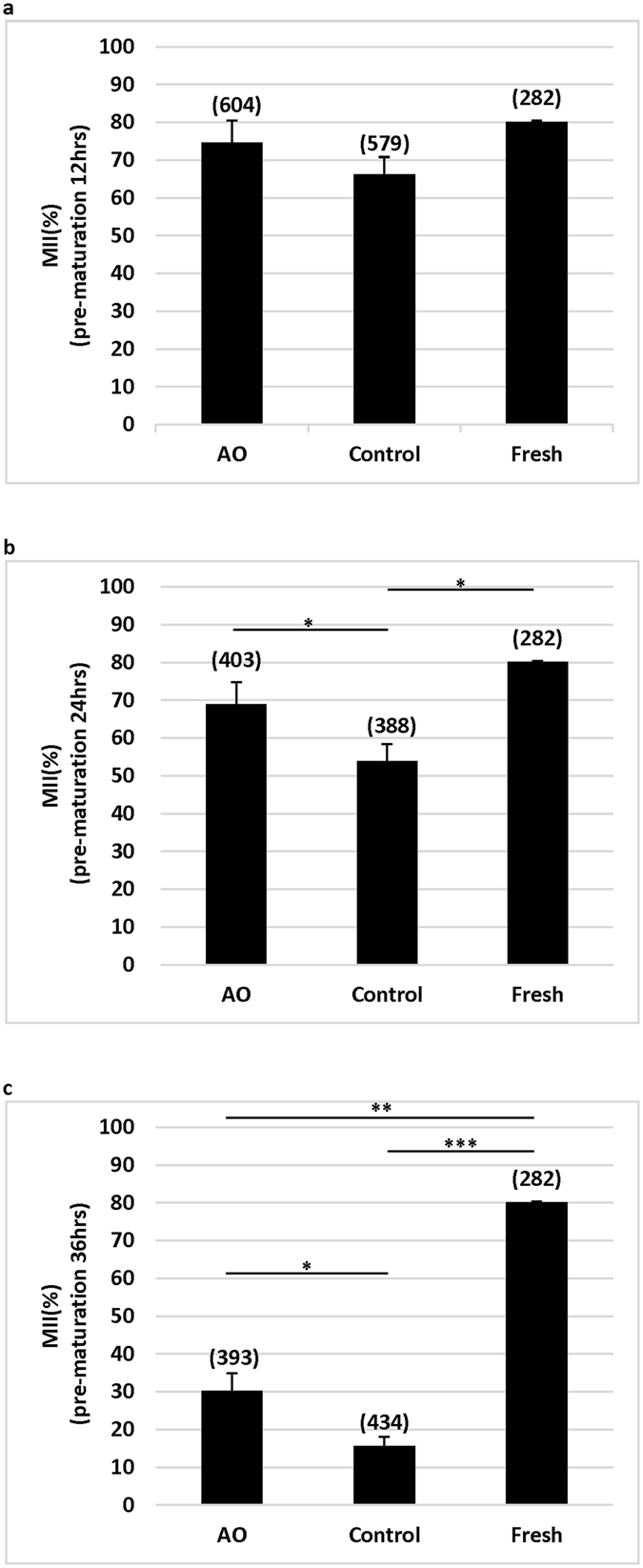
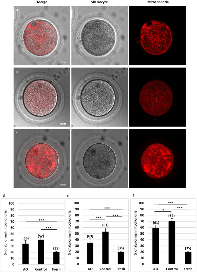
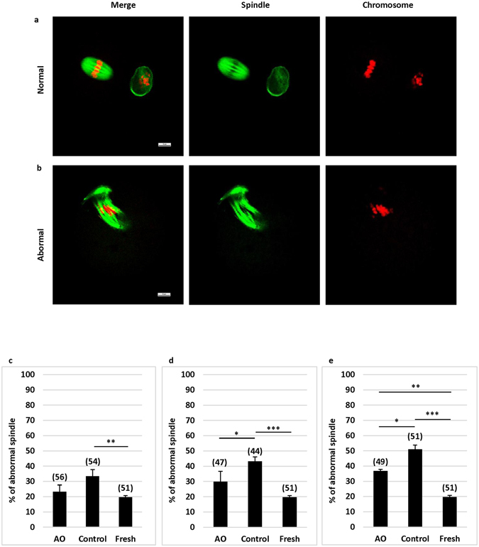
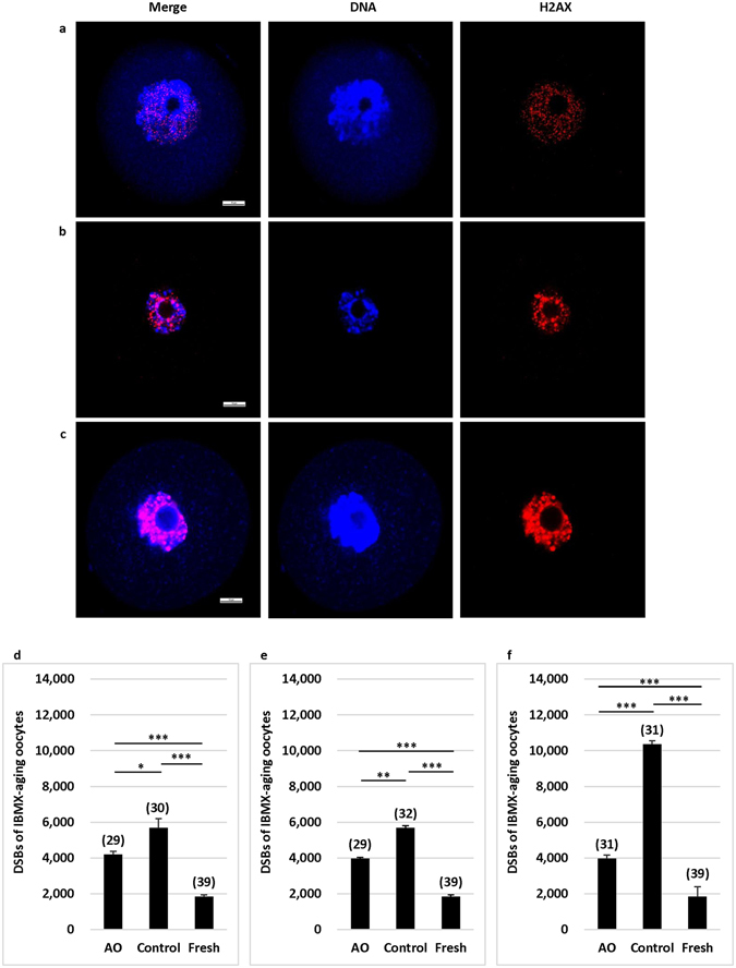
Pre-maturation aging of immature oocytes may adversely affect the fate of an oocyte. Oxidative stress is one of the most detrimental factors affecting oocyte developmental competence and maturation during aging. In this study, experiments were designed to examine whether supplementation of antioxidants in a culture medium could protect immature mouse oocytes from damages caused by oxidative stress. Mouse oocytes at germinal vesicle stage were prevented from meiosis resumption and cultured in a medium with or without antioxidants for 12-36 h to allow oocytes to undergo aging. After aging, oocytes were cultured for maturation. Nuclear maturation, mitochondria activity, spindle morphology and DNA integrity were examined after maturation. It was found that antioxidants had protective effects on the oocytes in terms of nuclear maturation, functional mitochondria, spindle morphology and DNA integrity. As aging time was prolonged from 12 to 36 h, the protective effect of antioxidants became more obvious. However, as compared with oocytes without aging, it was found that aging significantly inhibited nuclear maturation, impaired mitochondria function, and damaged the spindle and DNA. These results indicate that pre-maturation aging is detrimental to oocytes' competence to undergo maturation and other cellular activities, and antioxidants can protect oocytes from damages caused by aging.
Conflict of interest statement
The authors declare that they have no competing interests.
Figures
Similar articles
-
A pre-in vitro maturation medium containing cumulus oocyte complex ligand-receptor signaling molecules maintains meiotic arrest, supports the cumulus oocyte complex and improves oocyte developmental competence.Mol Hum Reprod. 2017 Sep 1;23(9):594-606. doi: 10.1093/molehr/gax032. Mol Hum Reprod. 2017. PMID: 28586460
-
N-acetyl-L-cysteine (NAC) delays post-ovulatory oocyte aging in mouse.Aging (Albany NY). 2019 Apr 12;11(7):2020-2030. doi: 10.18632/aging.101898. Aging (Albany NY). 2019. PMID: 30978175 Free PMC article.
-
l-carnitine supplementation during vitrification of mouse germinal vesicle stage-oocytes and their subsequent in vitro maturation improves meiotic spindle configuration and mitochondrial distribution in metaphase II oocytes.Hum Reprod. 2014 Oct 10;29(10):2256-68. doi: 10.1093/humrep/deu201. Epub 2014 Aug 11. Hum Reprod. 2014. PMID: 25113843
-
Delaying brain mitochondrial decay and aging with mitochondrial antioxidants and metabolites.Ann N Y Acad Sci. 2002 Apr;959:133-66. doi: 10.1111/j.1749-6632.2002.tb02090.x. Ann N Y Acad Sci. 2002. PMID: 11976193 Review.
-
Impact of Oxidative Stress on Age-Associated Decline in Oocyte Developmental Competence.Front Endocrinol (Lausanne). 2019 Nov 22;10:811. doi: 10.3389/fendo.2019.00811. eCollection 2019. Front Endocrinol (Lausanne). 2019. PMID: 31824426 Free PMC article. Review.
Cited by
-
Endogenous and Exogenous Modulation of Nrf2 Mediated Oxidative Stress Response in Bovine Granulosa Cells: Potential Implication for Ovarian Function.Int J Mol Sci. 2019 Apr 2;20(7):1635. doi: 10.3390/ijms20071635. Int J Mol Sci. 2019. PMID: 30986945 Free PMC article.
-
Beneficial effects of curcumin and capsaicin on cyclophosphamide-induced premature ovarian failure in a rat model.J Ovarian Res. 2018 Apr 26;11(1):33. doi: 10.1186/s13048-018-0409-9. J Ovarian Res. 2018. PMID: 29699594 Free PMC article.
-
Reprogramming of glucose metabolism of cumulus cells and oocytes and its therapeutic significance.Reprod Sci. 2022 Mar;29(3):653-667. doi: 10.1007/s43032-021-00505-6. Epub 2021 Mar 5. Reprod Sci. 2022. PMID: 33675030 Review.
-
Endometriosis Is a Cause of Infertility. Does Reactive Oxygen Damage to Gametes and Embryos Play a Key Role in the Pathogenesis of Infertility Caused by Endometriosis?Front Endocrinol (Lausanne). 2018 Nov 29;9:725. doi: 10.3389/fendo.2018.00725. eCollection 2018. Front Endocrinol (Lausanne). 2018. PMID: 30555421 Free PMC article. Review.
-
Role of N-acetylcysteine treatment in women with advanced age undergoing IVF/ICSI cycles: A prospective study.Front Med (Lausanne). 2022 Oct 4;9:917146. doi: 10.3389/fmed.2022.917146. eCollection 2022. Front Med (Lausanne). 2022. PMID: 36267623 Free PMC article.
References
Publication types
MeSH terms
Substances
LinkOut - more resources
Full Text Sources
Other Literature Sources
Medical
Research Materials
