. 2017 May 3;7(1):1457.
doi: 10.1038/s41598-017-01581-y.

Coaxial 3D bioprinting of self-assembled multicellular heterogeneous tumor fibers

Affiliations

Coaxial 3D bioprinting of self-assembled multicellular heterogeneous tumor fibers

Xingliang Dai et al. Sci Rep. .

Abstract

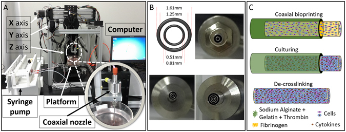
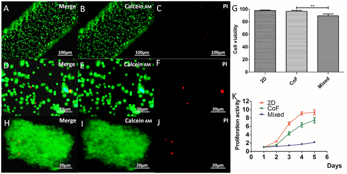
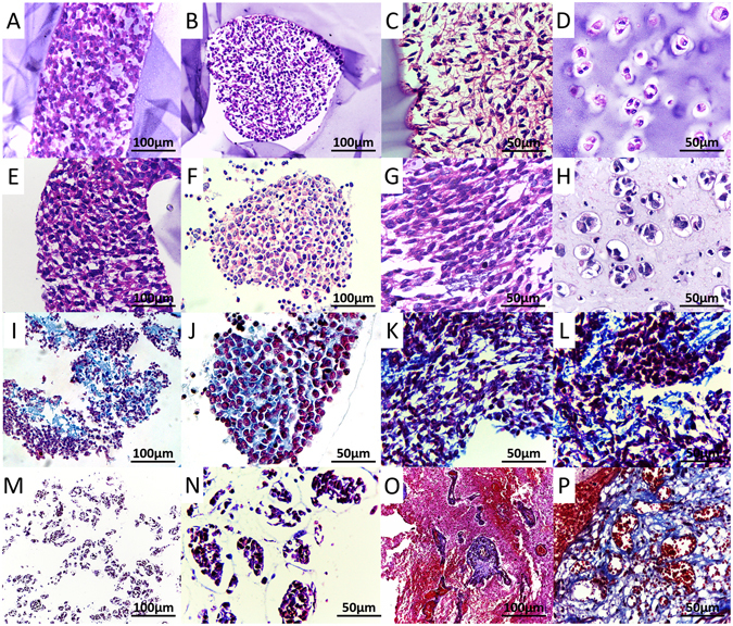
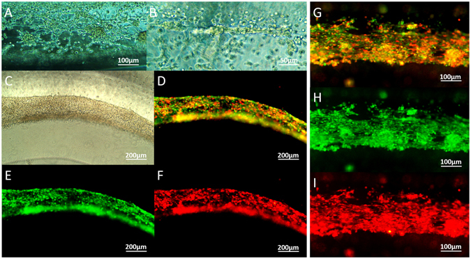
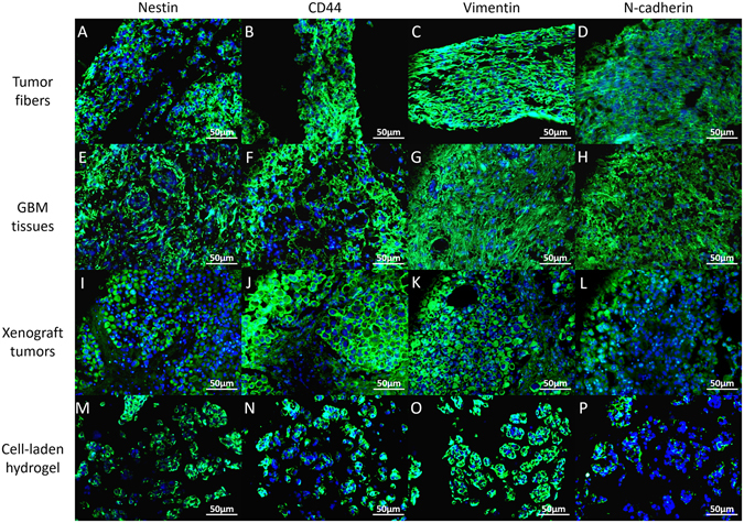
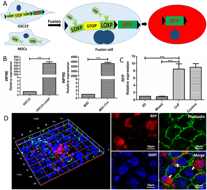
Three-dimensional (3D) bioprinting of living structures with cell-laden biomaterials has been achieved in vitro, however, some cell-cell interactions are limited by the existing hydrogel. To better mimic tumor microenvironment, self-assembled multicellular heterogeneous brain tumor fibers have been fabricated by a custom-made coaxial extrusion 3D bioprinting system, with high viability, proliferative activity and efficient tumor-stromal interactions. Therein, in order to further verify the sufficient interactions between tumor cells and stroma MSCs, CRE-LOXP switch gene system which contained GSCs transfected with "LOXP-STOP-LOXP-RFP" genes and MSCs transfected with "CRE recombinase" gene was used. Results showed that tumor-stroma cells interacted with each other and fused, the transcription of RFP was higher than that of 2D culture model and control group with cells mixed directly into alginate, respectively. RFP expression was observed only in the cell fibers but not in the control group under confocal microscope. In conclusion, coaxial 3D bioprinted multicellular self-assembled heterogeneous tumor tissue-like fibers provided preferable 3D models for studying tumor microenvironment in vitro, especially for tumor-stromal interactions.


Conflict of interest statement

The authors declare that they have no competing interests.

Figures

Figure 1
Figure 1
Coaxial 3D bioprinting system and the tumor fiber fabrication process. (A) Coaxial 3D bioprinting system contains a XYZ three-axis platform, a syringe pump, a coaxial nozzle and the computer-controlled system. (B) Different styles of the printing nozzles adapted with this bioprinting platform. (C) The schematic of the biofabrication process of multicellular heterogeneous tumor fiber: coaxial bioprinting, in vitro culturing and de-crosslinking.
Figure 2
Figure 2
Characteristics of the core-shell structures. (A and B) Integrity and continuity of the extruded liquid flow: the solution in the core stained with red or blue dyes can flow through the printed fiber shell without leaking out; (C) Permeability: Dye can uniformly diffuse out the structure within 40 minutes; D Layer by layer bioprinting (left 1–4) and perfusion of the printed construct with red dyes (right 1). Scale bars: (A) 40 mm, (B) 400 μm, (C) 20 mm, (D) 1 cm.
Figure 3
Figure 3
3D core-shell cell fibers. (A and B) GSC23 grew in stem cell medium (A) and complete medium (B); C: MSCs cultured in complete medium; D: Coaxial 3D bioprinted cell fibers; (E and F) Cell fiber with extrusion speed of 3 ml/h and 10 ml/h; (GL) Coaxial 3D bioprinted tumor fiber cultured for 1, 4, 7, 13, and 19 days. Scale bars: (AC) 50 μm, (D) 20 mm, (GK) 100 μm and (L) 400 μm.
Figure 4
Figure 4
Cell viability and proliferation. (AF) Live/dead assay for cell viability immediately after bioprinting; (G) Cell survival rate of CoF group comparing to cells without bioprinting; (HJ) Cell viability after culturing for 5 days; (K) Cell proliferation of CoF, 2D and mixed group after normalized to OD value of day 1. Scale bars: (AC) 100 μm, (DF) 20 μm, (H–J) 20 μm.
Figure 5
Figure 5
HE and Masson stainings. (AC) The coaxial 3D bioprinted tumor fiber within the alginate shell; (D) Cells mixed into alginate and cultured for 7 days; E-G: HE staining of the tumor fiber after removal of the alginate shell; H: Cells mixed into alginate and cultured for 21 days. (IL) Masson staining of the tumor fiber; (M,N) Masson staining of the cells mixed into alginate; (O,P) Masson staining of the GBM tissues. Scale bars: (A,B,E,F,I,M and O) 100 μm; (C,D,G,H,JL, N and P) 50 μm.
Figure 6
Figure 6
RFP/GFP traced tumor/stroma cell fibers. (A,B) Tumor cells and stroma cells fused into flakes and strands on day 2; (CF) Cell fibers containing RFP-expressing tumor cells and GFP-expressing MSCs after bioprinting and cultured in vitro for 3 days; (GI) Cell fibers cultured in vitro for 7 days. Scale bars: (A and GI) 100 μm; (B) 50 μm; (CF) 200 μm.
Figure 7
Figure 7
Immunohistology. Expression of biomarkers (Nestin, CD44, Vimentin and N-cadherin) in coaxial bioprinted tumor fibers, comparing to GBM tissues, xenograft tumors and cell-laden hydrogel (cells mixed into A/G). Scale bars: 50 μm.
Figure 8
Figure 8
Cell fusion. (A) Schematic of interaction principle between CRE enzyme and LOXP-STOP- LOXP –RFP gene; (B) qRT-PCR validation of the transfection efficiency in GSC23 and MSC cells; (C) qRT-PCR validation of the RFP expression in CoF, comparing to 2D group, mixed group and coaxial group without adding fibrinogen to cell suspension (control); (D) Confocal images of GSC23-MSCs fibers, red is RFP (white arrow), phalloidin was used to stain the cytoskeletal structures (green) and DAPI was used to stain the cell nucleus (blue). Scale bars: (D) 10 μm.

Similar articles

Cited by

References

    1. Murphy SV, Atala A. 3D bioprinting of tissues and organs. Nat Biotechnol. 2014;32:773–785. doi: 10.1038/nbt.2958. - DOI - PubMed
    1. Li J, Chen M, Fan X, Zhou H. Recent advances in bioprinting techniques: approaches, applications and future prospects. J Transl Med. 2016;20:271. doi: 10.1186/s12967-016-1028-0. - DOI - PMC - PubMed
    1. Murphy SV, Skardal A, Atala A. Evaluation of hydrogels for bio-printing applications. J Biomed Mater Res A. 2013;101:272–84. doi: 10.1002/jbm.a.34326. - DOI - PubMed
    1. Knowlton S, Onal S, Yu CH, Zhao JJ, Tasoglu S. Bioprinting for cancer research. Trends Biotechnol. 2015;33:504–13. doi: 10.1016/j.tibtech.2015.06.007. - DOI - PubMed
    1. Ma Y, et al. Bioprinting 3D cell laden hydrogel microarray for screening human periodontal ligament stem cell response to extracellular matrix. Biofabrication. 2015;7:044105. doi: 10.1088/1758-5090/7/4/044105. - DOI - PubMed

Publication types

MeSH terms