

Construction of a highly efficient CRISPR/Cas9-mediated duck enteritis virus-based vaccine against H5N1 avian influenza virus and duck Tembusu virus infection
- PMID: 28469192
- PMCID: PMC5431151
- DOI: 10.1038/s41598-017-01554-1
Construction of a highly efficient CRISPR/Cas9-mediated duck enteritis virus-based vaccine against H5N1 avian influenza virus and duck Tembusu virus infection
Abstract
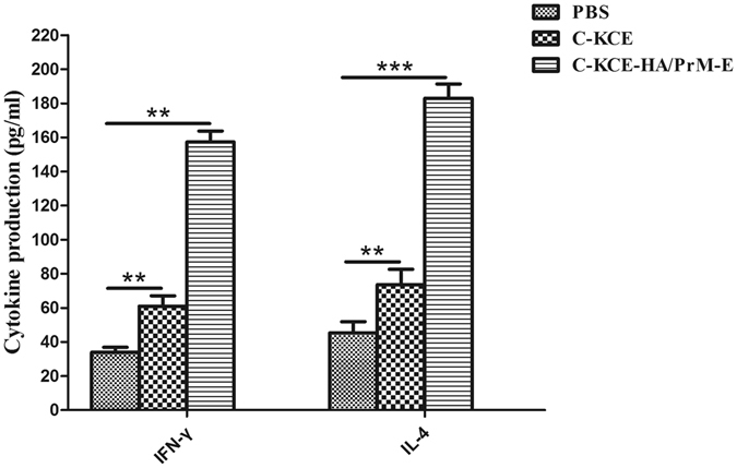
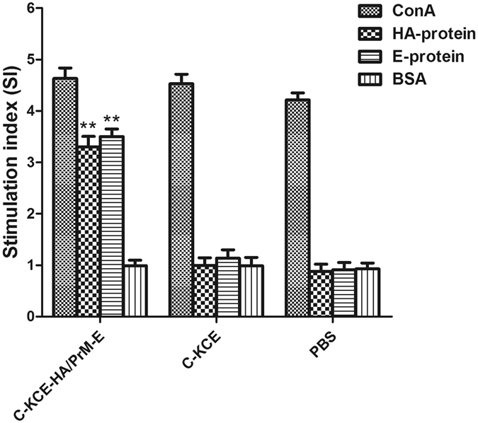
Duck enteritis virus (DEV), duck tembusu virus (DTMUV), and highly pathogenic avian influenza virus (HPAIV) H5N1 are the most important viral pathogens in ducks, as they cause significant economic losses in the duck industry. Development of a novel vaccine simultaneously effective against these three viruses is the most economical method for reducing losses. In the present study, by utilizing a clustered regularly interspaced short palindromic repeats (CRISPR)/associated 9 (Cas9)-mediated gene editing strategy, we efficiently generated DEV recombinants (C-KCE-HA/PrM-E) that simultaneously encode the hemagglutinin (HA) gene of HPAIV H5N1 and pre-membrane proteins (PrM), as well as the envelope glycoprotein (E) gene of DTMUV, and its potential as a trivalent vaccine was also evaluated. Ducks immunized with C-KCE-HA/PrM-E enhanced both humoral and cell-mediated immune responses to H5N1 and DTMUV. Importantly, a single-dose of C-KCE-HA/PrM-E conferred solid protection against virulent H5N1, DTMUV, and DEV challenges. In conclusion, these results demonstrated for the first time that the CRISPR/Cas9 system can be applied for modification of the DEV genome rapidly and efficiently, and that recombinant C-KCE-HA/PrM-E can serve as a potential candidate trivalent vaccine to prevent H5N1, DTMUV, and DEV infections in ducks.
Conflict of interest statement
The authors declare that they have no competing interests.
Figures
Similar articles
-
Efficient strategy for constructing duck enteritis virus-based live attenuated vaccine against homologous and heterologous H5N1 avian influenza virus and duck enteritis virus infection.Vet Res. 2015 Apr 16;46(1):42. doi: 10.1186/s13567-015-0174-3. Vet Res. 2015. PMID: 25889564 Free PMC article.
-
Construction of a recombinant duck enteritis virus (DEV) expressing hemagglutinin of H5N1 avian influenza virus based on an infectious clone of DEV vaccine strain and evaluation of its efficacy in ducks and chickens.Virol J. 2015 Aug 13;12:126. doi: 10.1186/s12985-015-0354-9. Virol J. 2015. PMID: 26263920 Free PMC article.
-
Efficient strategy to generate a vectored duck enteritis virus delivering envelope of duck Tembusu virus.Viruses. 2014 Jun 20;6(6):2428-43. doi: 10.3390/v6062428. Viruses. 2014. PMID: 24956180 Free PMC article.
-
Vaccination of gallinaceous poultry for H5N1 highly pathogenic avian influenza: current questions and new technology.Virus Res. 2013 Dec 5;178(1):121-32. doi: 10.1016/j.virusres.2013.03.004. Epub 2013 Mar 21. Virus Res. 2013. PMID: 23524326 Review.
-
Advancements in Research on Duck Tembusu Virus Infections.Viruses. 2024 May 20;16(5):811. doi: 10.3390/v16050811. Viruses. 2024. PMID: 38793692 Free PMC article. Review.
Cited by
-
Assembly-defective Tembusu virus ectopically expressing capsid protein is an approach for live-attenuated flavivirus vaccine development.NPJ Vaccines. 2022 May 12;7(1):51. doi: 10.1038/s41541-022-00468-y. NPJ Vaccines. 2022. PMID: 35550523 Free PMC article.
-
CRISPR/Cas9-Advancing Orthopoxvirus Genome Editing for Vaccine and Vector Development.Viruses. 2018 Jan 22;10(1):50. doi: 10.3390/v10010050. Viruses. 2018. PMID: 29361752 Free PMC article. Review.
-
CRISPR/Cas9-edited duck enteritis virus expressing Pmp17G of Chlamydia psittaci induced protective immunity in ducklings.Pathog Dis. 2024 Feb 7;82:ftae027. doi: 10.1093/femspd/ftae027. Pathog Dis. 2024. PMID: 39400699 Free PMC article.
-
The Neutralizing Antibody Response Elicited by Tembusu Virus Is Affected Dramatically by a Single Mutation in the Stem Region of the Envelope Protein.Front Microbiol. 2020 Oct 22;11:585194. doi: 10.3389/fmicb.2020.585194. eCollection 2020. Front Microbiol. 2020. PMID: 33193231 Free PMC article.
-
Latest Advances of Virology Research Using CRISPR/Cas9-Based Gene-Editing Technology and Its Application to Vaccine Development.Viruses. 2021 Apr 28;13(5):779. doi: 10.3390/v13050779. Viruses. 2021. PMID: 33924851 Free PMC article. Review.
References
-
- Newcomb SS. Duck virus enteritis (duck plague) epizootiology and related investigations. Journal of the American Veterinary Medical Association. 1968;153:1897–1902. - PubMed
Publication types
MeSH terms
Substances
Supplementary concepts
LinkOut - more resources
Full Text Sources
Other Literature Sources
Medical
Research Materials
