. 2017 May 3;7(1):1416.
doi: 10.1038/s41598-017-01641-3.

A HAND to TBX5 Explains the Link Between Thalidomide and Cardiac Diseases

Affiliations

A HAND to TBX5 Explains the Link Between Thalidomide and Cardiac Diseases

Athar Khalil et al. Sci Rep. .

Abstract

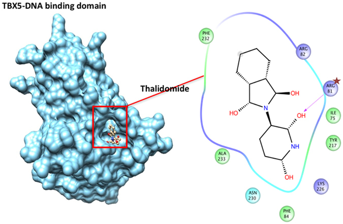
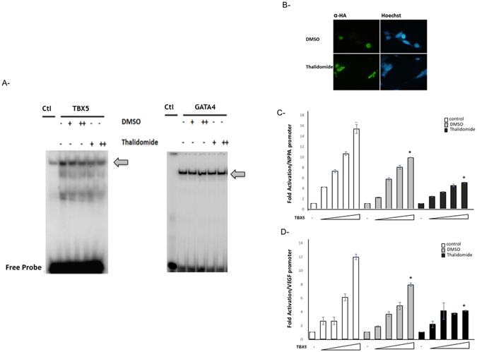
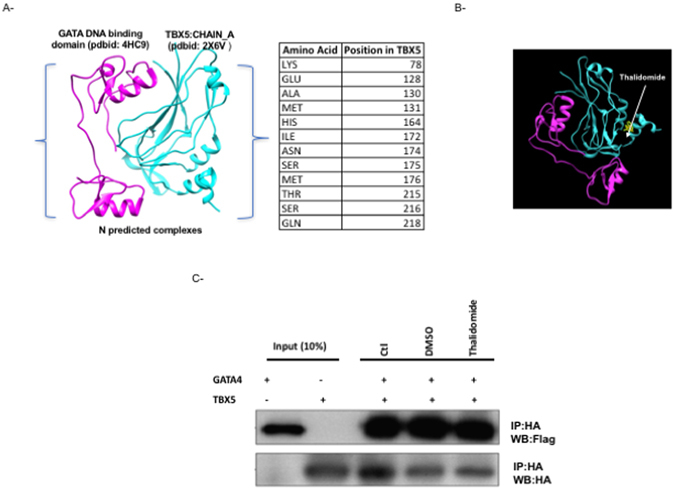
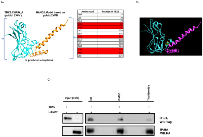
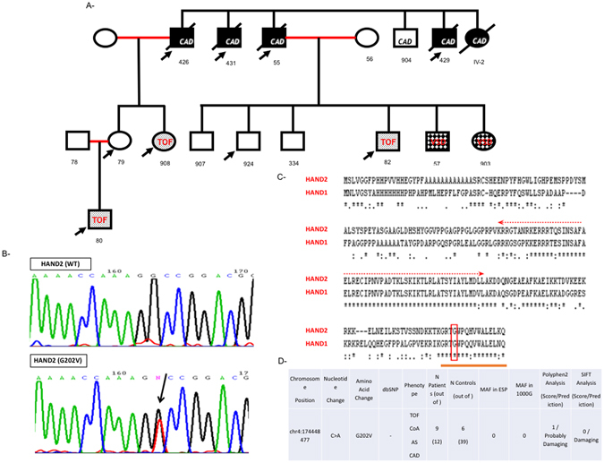
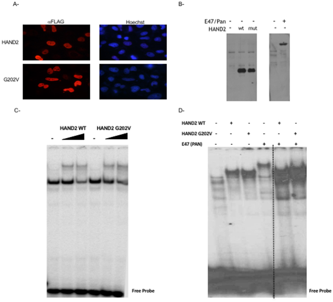
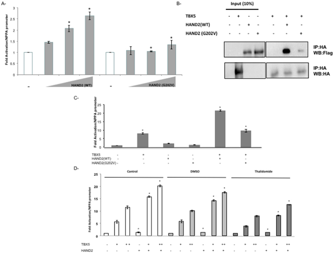
Congenital heart disease is the leading cause of death in the first year of life. Mutations only in few genes have been linked to some cases of CHD. Thalidomide was used by pregnant women for morning sickness but was removed from the market because it caused severe malformations including CHDs. We used both in silico docking software, and in vitro molecular and biochemical methods to document a novel interaction involving Thalidomide, TBX5, and HAND2. Thalidomide binds readily to TBX5 through amino acids R81, R82, and K226 all implicated in DNA binding. It reduces TBX5 binding to DNA by 40%, and suppresses TBX5 mediated activation of the NPPA and VEGF promoters by 70%. We documented a novel interaction between TBX5 and HAND2, and showed that a p.G202V HAND2 variant associated with CHD and coronary artery diseases found in a large Lebanese family with high consanguinity, drastically inhibited this interaction by 90%. Similarly, thalidomide inhibited the TBX5/HAND2 physical interaction, and the in silico docking revealed that the same amino acids involved in the interaction of TBX5 with DNA are also involved in its binding to HAND2. Our results establish a HAND2/TBX5 pathway implicated in heart development and diseases.


Conflict of interest statement

The authors declare that they have no competing interests.

Figures

Figure 1
Figure 1
Thalidomide specific binding to the T-box domain of TBX5. The corresponding protein-ligand interaction diagram shows the most relevant amino acid within 5 Angstroms of thalidomide. Arginine at position 81 (arrow) is proposed as the main player for the TBX5-thalidomide docking simulation. The T-box domain of TBX5 (pdbid: 2X6V) was retrieved from the Protein Database (PDB). All colored amino acids with their positions are the ones in close proximity with Thalidomide.
Figure 2
Figure 2
Thalidomide specific inhibition of TBX5 binding and activity. Interactions of TBX5 and GATA4 with the NPPA TBE and GATA sites was assessed by electrophoretic mobility shift assays using nuclear extracts from HEK293T cells overexpressing HA-TBX5 or Flag-GATA4 (A). The binding of TBX5 (arrow) is suppressed by 42% in the presence of 4 µM of thalidomide, while that of GATA4 (arrow) was not affected. (+2 µM, ++4 µM). DMSO treatment was used as a negative control. Immunofluorescence of JR1 cells transfected with the plasmid overexpressing a HA-tagged TBX5 protein and then treated with either 4 µM DMSO or thalidomide. The localization of TBX5 was visualized using an anti‐ HA antibody followed by a fluorescent secondary antibody (B). Nuclei of cells were visualized using the Hoechst dye (blue color). TBX5 showed nuclear localization (green color) in both conditions. (Magnification X40). Same results were obtained for Hela and Hek293 cells. Transcriptional activity was assessed by luciferase assay. Increasing doses of TBX5 (100 to 500 ng) were transiently co-transfected with the rat NPPA (C), or mouse VEGF (D) promoters coupled to luciferase in HEK293 cells. Treatment with thalidomide (4 µM) or its equivalent amount of DMSO was done after 24 hours, and cells were harvested for luciferase assay. Relative luciferase activities are represented as fold changes. The data are the means of 3 independent experiments done in duplicates ± SE. Significance (*P < 0.05) was assessed using the one‐way ANOVA test.
Figure 3
Figure 3
GATA4/TBX5 interaction is not affected by Thalidomide. Protein-protein docking interface between TBX5 and GATA4 modeled DNA binding domain as retrieved from PDB. Only Residues of TBX5 within 5 angstroms of this interaction are shown (A). The position of thalidomide in the context of the predicted TBX5-GATA4 interaction is clearly away from the interface region (B): TBX5 (cyan) and GATA4 (magenta). The physical interaction between both proteins was assessed by co-immunoprecipitation in the presence or absence (−) of either 4 µM thalidomide or DMSO (C). Ten times the quantity of proteins loaded for western blot was used for immune-precipitation. Nuclear lysates for both GATA4 and TBX5 were immune-precipitated with anti‐HA antibody and GATA4 proteins were visualized by western blot with an anti‐flag antibody. Membrane stripping and subsequent western blot analysis was performed with anti‐HA antibody in order to detect TBX5 proteins. Quantitation of the bands didn’t show any significant change. (Ctr: control, WB: Western-Blot, IP: Immuno-Precipitation).
Figure 4
Figure 4
HAND2/TBX5 interaction drastically inhibited by Thalidomide. Protein-protein docking interface between TBX5 and HAND2 modeled DNA binding domain as retrieved from PDB. Only Residues of TBX5 within 5 angstroms of this interaction are shown (A). Those in red are associated with both TBX5-DNA and TBX5-HAND2 interaction from modeling simulation, which shows the position of thalidomide in the context of predicted TBX5-HAND2 interaction (B): TBX5 (cyan) and HAND2 (magenta). The physical interaction between both proteins was assessed by co-immunoprecipitation in the presence or absence (−) of either 4 µM thalidomide or DMSO (C). Ten times the quantity of proteins loaded for western blot was used for immunoprecipitation. Nuclear lysates of both HAND2 and TBX5 were immunoprecipitated with anti‐HA antibody and HAND2 proteins were visualized by western blot with and anti‐flag antibody. Membrane stripping and subsequent western blot analysis was performed with anti‐HA antibody in order to detect TBX5 proteins. Quantitation of the bands showed more than 80% decrease in the interaction when the extracts were incubated in the presence of Thalidomide. (Ctr: control, WB: immunoblot, IP: immnuoprecipitation).
Figure 5
Figure 5
Phenotype/Genotype of the p.G202V HAND2 within the affected family. The pedigree (A) was annotated using the clear squares and circle for healthy males and females in the family. The marriages between first-degree cousins were annotated as red lines, and the affected individuals with the appropriate symbols. The p.G202V variant was indicated by arrow on all affected individuals. All deceased individuals were annotated with black strikethrough squares or circles. Numbers underneath each refer to the code given for each DNA sample corresponding to the individual. (CAD: coronary artery disease, TOF: tetralogy of Fallot, VSD: ventricular septal defect). Chromatograms (B) showing Sanger sequencing results for WT and the p.G202V variant. The boxed region corresponds to the heterozygous C > A nucleotide change read with the reverse primer. Sequence conservation of the G202 amino acid at position 202 between the HAND proteins (HAND1 and 2) using the clustalw tool (www.ebi.ac.uk/Tools/msa/clustalw2/). The Glycine residue is boxed within the highly conserved C-terminal domain while the conserved bHLH domain is marked with an arrow (C). The genotype and phenotype of the extended family members are listed along the minor allele frequency (MAF) and the in silico predictions of the mutation (D).
Figure 6
Figure 6
Characterization of the p.G202V protein in vitro. Transiently transfected Hela cells with plasmids harboring either the wild type HAND2 or the p.G202V variant were fixed and stained with an anti-Flag antibody. The secondary fluorescent antibody showed no change in the nuclear staining (in red) for both proteins as compared to the Hoechst staining (Magnification X40) (A). Western blot using the same anti-Flag antibody shows equal expression of both the wild type (wt) and mutated (mut) proteins in Hek293 cells, as well as the transiently overexpressed E47/Pan protein (B). Gel Shift analyses with increasing doses from the same nuclear extracts as in (C) were used on a consensus HAND binding site showed similar pattern of binding for both the wild type and mutant HAND2 proteins (D). The same pattern was observed when mixing the same amount of the HAND2 proteins with E47/Pan. The panel was separated by dotted lines to highlight the fact that the presented figure is a merge of two different gels run simultaneously.
Figure 7
Figure 7
The TBX5/HAND2 Functional Interaction Inhibited both by the p.G202V variant and Thalidomide. The transcriptional activity of HAND2 was assessed in co-transfection assays using the rat NPPA promoter coupled to luciferase which showed a drastic decrease in the p.G202V mutant ability to activate its target (A). Relative luciferase activities are represented as fold changes. The data are the means of 3 independent experiments done in duplicates ± SE. Significance (*P < 0.05) was assessed using the one‐way ANOVA test. HA‐tagged TBX5 and Flag‐tagged HAND2 (WT and G202V) proteins were extracted from transiently transfected HEK293 cells. Nuclear lysates for both HAND2 and TBX5 proteins were immune-precipitated with anti‐HA antibody, and visualized with western blot via anti‐flag antibody. Membrane stripping and subsequent western blot analysis was performed with anti‐HA antibody in order to detect TBX5 proteins. Ten times the quantity of proteins loaded for western blot was used for immune-precipitation. Quantitation of the bands shows up to 80% inhibition of the interaction when the HAND2 mutated protein is used instead of the wild type protein (B). Transcriptional activity was assessed by luciferase assay (C,D). TBX5 and HAND2 (WT or G202V) were transiently co-transfected with the rat NPPA promoter coupled to luciferase in HEK293 cells (C), then in the presence of Thalidomide (D). Relative luciferase activities are represented as fold changes. The data are the means of 3 independent experiments done in duplicates ± SE. Significance (*P < 0.05) was assessed using the one‐way ANOVA test. (+) is for 300 ng of DNA, and (++) for 700 ng of DNA. The increasing doses start at 100 reaching up to 500 ng.

Similar articles

Cited by

References

    1. Clowes C, et al. The functional diversity of essential genes required for mammalian cardiac development. Genesis. 2014;52:713–737. doi: 10.1002/dvg.22794. - DOI - PMC - PubMed
    1. Srivastava D, Olson EN. A genetic blueprint for cardiac development. Nature. 2000;407:221–226. doi: 10.1038/35025190. - DOI - PubMed
    1. Christoffels VM, et al. Chamber formation and morphogenesis in the developing mammalian heart. Dev Biol. 2000;223:266–278. doi: 10.1006/dbio.2000.9753. - DOI - PubMed
    1. Lin Q, Srivastava D, Olson EN. A transcriptional pathway for cardiac development. Cold Spring Harb Symp Quant Biol. 1997;62:405–411. doi: 10.1101/SQB.1997.062.01.048. - DOI - PubMed
    1. Olson EN, Srivastava D. Molecular pathways controlling heart development. Science. 1996;272:671–676. doi: 10.1126/science.272.5262.671. - DOI - PubMed

Publication types

MeSH terms

LinkOut - more resources