

A Case Report of Krukenbergs Tumor with Cutaneous Seeding
- PMID: 28469341
- PMCID: PMC5398111
- DOI: 10.4103/0971-5851.203504
A Case Report of Krukenbergs Tumor with Cutaneous Seeding
Abstract
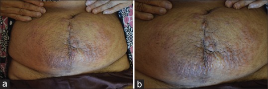
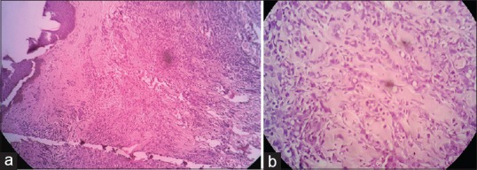
A 47-year-old female patient presented with painless skin colored and erythematous papules coalesced to form plaques over lower abdomen for 10 days. She had undergone exploratory laparotomy with hysterectomy and bilateral oophorectomy 1 month ago, and histopathology was reported as Krukenbergs tumor. She was getting evaluated for primary, when she was referred to dermatology. A clinical diagnosis of cutaneous infiltration of tumor was made, and biopsy was done from a representative lesion which showed features suggestive of metastatic poorly differentiated adenocarcinoma. In the majority of cases in the past, cutaneous metastasis is seen much later in the course of the disease. High degree of suspicion and histopathology was helpful in the diagnosis of underlying malignancy in our patient.
Keywords: Cutaneous metastasis; Krukenbergs tumor; poorly differentiated adenocarcinoma.
Conflict of interest statement
There are no conflicts of interest.
Figures
Similar articles
-
Folliculotropic Cutaneous Metastases and Lymphangitis Carcinomatosa: When Cutaneous Metastases of Breast Carcinoma Are Mistaken for Cutaneous Infections.Acta Dermatovenerol Croat. 2016 Jun;24(2):154-7. Acta Dermatovenerol Croat. 2016. PMID: 27477179
-
Sister Mary Joseph's nodule as a presenting sign of internal malignancy.Skinmed. 2006 Sep-Oct;5(5):256-8. doi: 10.1111/j.1540-9740.2006.04826.x. Skinmed. 2006. PMID: 16957443
-
Cytokeratin 7-positive/cytokeratin 20-negative cecal adenocarcinoma metastatic to the uterine cervix: a case report.World J Surg Oncol. 2016 Jan 25;14(1):22. doi: 10.1186/s12957-016-0774-z. World J Surg Oncol. 2016. PMID: 26810414 Free PMC article.
-
Metachronous Ovarian Metastases in a Patient with Primary Colorectal Cancer. A Case Report and Review of the Literature.Am J Case Rep. 2019 Oct 15;20:1515-1520. doi: 10.12659/AJCR.917957. Am J Case Rep. 2019. PMID: 31611546 Free PMC article. Review.
-
Palliative Chemotherapy: Does It Only Provide False Hope? The Role of Palliative Care in a Young Patient With Newly Diagnosed Metastatic Adenocarcinoma.J Adv Pract Oncol. 2017 May-Jun;8(4):382-386. Epub 2017 May 1. J Adv Pract Oncol. 2017. PMID: 30018843 Free PMC article. Review.
References
-
- Rao R, Balachandran C, Rao L. Zosteriform cutaneous metastases: A case report and brief review of literature. Indian J Dermatol Venereol Leprol. 2010;76:447. - PubMed
-
- Rendi MH, Dhar AD. Cutaneous metastasis of rectal adenocarcinoma. Dermatol Nurs. 2003;15:131–2. - PubMed
-
- Sarid D, Wigler N, Gutkin Z, Merimsky O, Leider-Trejo L, Ron IG. Cutaneous and subcutaneous metastases of rectal cancer. Int J Clin Oncol. 2004;9:202–5. - PubMed
-
- Hussein MR. Skin metastasis: A pathologist's perspective. J Cutan Pathol. 2010;37:e1–20. - PubMed
-
- Riley S, Wah T. Cutaneous metastasis of esophageal adenocarcinoma with an unusual presentation. J Clin Ultrasound. 2007;35:289–92. - PubMed
Publication types
LinkOut - more resources
Full Text Sources
Other Literature Sources
Miscellaneous
