Magnetic Resonance Imaging Detection of Intraplaque Hemorrhage
- PMID: 28469441
- PMCID: PMC5348123
- DOI: 10.1177/1178623X17694150
Magnetic Resonance Imaging Detection of Intraplaque Hemorrhage
Abstract
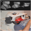
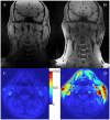
Carotid artery atherosclerosis is a major cause of ischemic stroke. For more than 30 years, future stroke risk and carotid stroke etiology have been determined using percent diameter stenosis based on clinical trials in the 1990s. In the past 10 years, magnetic resonance imaging (MRI) sequences have been developed to detect carotid intraplaque hemorrhage. By detecting carotid intraplaque hemorrhage, MRI identifies potential stroke sources that are often overlooked by lumen imaging. In addition, MRI can dramatically improve assessment of future stroke risk beyond lumen stenosis alone. In this review, we discuss the use of heavily T1-weighted MRI sequences used to detect carotid intraplaque hemorrhage. In addition, advances in ciné imaging, motion robust techniques, and specialized neck coils will be reviewed. Finally, the clinical use and future impact of MRI plaque hemorrhage imaging will be discussed.
Keywords: MRI; atherosclerosis; intraplaque hemorrhage; stroke.
Conflict of interest statement
DECLARATION OF CONFLICTING INTERESTS: The author(s) declared no potential conflicts of interest with respect to the research, authorship, and/or publication of this article. Disclosures and Ethics As a requirement of publication, author(s) have provided to the publisher signed confirmation of compliance with legal and ethical obligations including, but not limited to, the following: authorship and contributorship, conflicts of interest, privacy and confidentiality, and (where applicable) protection of human and animal research subjects. The authors have read and confirmed their agreement with the ICMJE authorship and conflict of interest criteria. The authors have also confirmed that this article is unique and not under consideration or published in any other publication, and that they have permission from rights holders to reproduce any copyrighted material. Any disclosures are made in this section. The external blind peer reviewers report no conflicts of interest.
Figures


References
-
- Mozaffarian D, Benjamin EJ, Go AS, et al. Heart disease and stroke statistics-2015 update: a report from the American Heart Association. Circulation. 2015;131:e29–e322. - PubMed
-
- Clinical alert: benefit of carotid endarterectomy for patients with high-grade stenosis of the internal carotid artery. National Institute of Neurological Disorders and Stroke Stroke and Trauma Division. North American Symptomatic Carotid Endarterectomy Trial (NASCET) investigators. Stroke. 1991;22:816–817. - PubMed
-
- Endarterectomy for asymptomatic carotid artery stenosis. Executive Committee for the Asymptomatic Carotid Atherosclerosis Study. JAMA. 1995;273:1421–1428. - PubMed
-
- Altaf N, Beech A, Goode SD, et al. Carotid intraplaque hemorrhage detected by magnetic resonance imaging predicts embolization during carotid endarterectomy. J Vasc Surg. 2007;46:31–36. - PubMed
Publication types
Grants and funding
LinkOut - more resources
Full Text Sources
Other Literature Sources
Medical
Miscellaneous

