

Ultrasound-Diagnosed Tibia Stress Fracture: A Case Report
- PMID: 28469488
- PMCID: PMC5390922
- DOI: 10.1177/1179544117702866
Ultrasound-Diagnosed Tibia Stress Fracture: A Case Report
Abstract
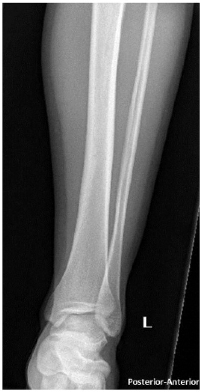
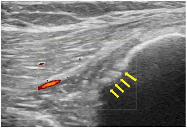
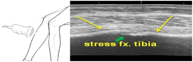
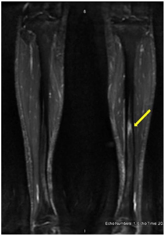
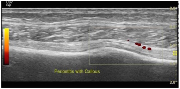
Stress fractures are a frequent cause of lower extremity pain in athletes, and especially in runners. Plain imaging has a low sensitivity. Magnetic resonance imaging (MRI) or bone scan scintigraphy is the criterion standard, but expensive. We present the case of a young female distance runner with left shin pain. Plain radiography was unremarkable. Ultrasound showed focal hyperechoic elevation of the periosteum with irregularity over the distal tibia and increased flow on Doppler. These findings were consistent with a distal tibia stress fracture and confirmed by MRI. Examination of our case will highlight the utility of considering an ultrasound for diagnosis of tibial stress fracture.
Keywords: Stress fracture; tibia; ultrasound.
Conflict of interest statement
DECLARATION OF CONFLICTING INTERESTS: The author(s) declared no potential conflicts of interest with respect to the research, authorship, and/or publication of this article.
Figures
References
-
- Dobrindt O, Hoffmeyer B, Ruf J, et al. Blinded-read of bone scintigraphy: the impact on diagnosis and healing time for stress injuries with emphasis on the foot. Clin Nucl Med. 2011;36:186–191. - PubMed
-
- Papalada A, Malliaropoulos N, Tsitas K, et al. Ultrasound as a primary evaluation tool of bone stress injuries in elite track and field athletes. Am J Sports Med. 2012;40:915–919. - PubMed
-
- Reeder MT, Dick BH, Atkins JK, Pribis AB, Martinez JM. Stress fractures: current concepts of diagnosis and treatment. Sports Med. 1996;22:198–212. - PubMed
-
- Geslien GE, Thrall JH, Espinosa JL, et al. Early detection of stress fractures using 99mTC-polyphosphonate. Radiol. 1976;121:683–687. - PubMed
-
- Bianchi S, Luong DH. Stress fractures of the ankle malleoli diagnosed by ultrasound: a report of 6 cases. Skeletal Radiol. 2014;43:813–818. - PubMed
Publication types
LinkOut - more resources
Full Text Sources
Other Literature Sources
