Unmasking the Role of Uptake Transporters for Digoxin Uptake Across the Barriers of the Central Nervous System in Rat
- PMID: 28469522
- PMCID: PMC5392048
- DOI: 10.1177/1179573517693596
Unmasking the Role of Uptake Transporters for Digoxin Uptake Across the Barriers of the Central Nervous System in Rat
Abstract
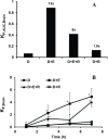
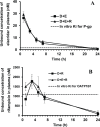
The role of uptake transporter (organic anion-transporting polypeptide [Oatp]) in the disposition of a P-glycoprotein (P-gp) substrate (digoxin) at the barriers of central nervous system, namely, the blood-brain barrier (BBB), blood-spinal cord barrier (BSCB), and brain-cerebrospinal fluid barrier (BCSFB), was studied using rat as a preclinical species. In vivo chemical inhibition of P-gp and Oatp was achieved using elacridar and rifampicin, respectively. Our findings show that (1) digoxin had a low brain-to-plasma concentration ratio (B/P) (0.07) in rat; (2) in the presence of elacridar, the B/P of digoxin increased by about 12-fold; (3) rifampicin administration alone did not change the digoxin B/P significantly when compared with digoxin B/P alone; (4) rifampicin administration along with elacridar resulted only in 6-fold increase in the B/P of digoxin; (5) similar fold changes and trends were seen with the spinal cord-to-plasma concentration ratio of digoxin, indicating the similarity between BBB and the BSCB; and (6) unlike BBB and BSCB, the presence of rifampicin further increased the cerebrospinal fluid-to-plasma concentration ratio (CSF/P) for digoxin, suggesting a differential orientation of the uptake transporters at the BCSFB (CSF to blood) compared with the BBB (blood to brain). The observations for digoxin uptake, at least at the BBB and the BSCB, advocate the importance of uptake transporters (Oatps). However, the activity of such uptake transporters became evident only after inhibition of the efflux transporter (P-gp).
Keywords: CNS; Digoxin; P-glycoprotein; blood-brain barrier; blood-cerebrospinal fluid barrier; blood-spinal cord barrier; organic anion transporting polypeptide.
Conflict of interest statement
DECLARATION OF CONFLICTING INTERESTS: The author(s) declared no potential conflicts of interest with respect to the research, authorship, and/or publication of this article.
Figures


Similar articles
-
Estimation of the unbound brain concentration of P-glycoprotein substrates or nonsubstrates by a serial cerebrospinal fluid sampling technique in rats.Mol Pharm. 2014 Feb 3;11(2):477-85. doi: 10.1021/mp400436d. Epub 2014 Jan 9. Mol Pharm. 2014. PMID: 24380373
-
Organic Anion-Transporting Polypeptide 1a4 (Oatp1a4/Slco1a4) at the Blood-Arachnoid Barrier is the Major Pathway of Sulforhodamine-101 Clearance from Cerebrospinal Fluid of Rats.Mol Pharm. 2019 May 6;16(5):2021-2027. doi: 10.1021/acs.molpharmaceut.9b00005. Epub 2019 Apr 22. Mol Pharm. 2019. PMID: 30977661
-
Effect of transporter inhibition on the distribution of cefadroxil in rat brain.Fluids Barriers CNS. 2014 Nov 14;11(1):25. doi: 10.1186/2045-8118-11-25. eCollection 2014. Fluids Barriers CNS. 2014. PMID: 25414790 Free PMC article.
-
Functional Expression of P-glycoprotein and Organic Anion Transporting Polypeptides at the Blood-Brain Barrier: Understanding Transport Mechanisms for Improved CNS Drug Delivery?AAPS J. 2017 Jul;19(4):931-939. doi: 10.1208/s12248-017-0081-9. Epub 2017 Apr 26. AAPS J. 2017. PMID: 28447295 Free PMC article. Review.
-
Potential role of ABC transporters as a detoxification system at the blood-CSF barrier.Adv Drug Deliv Rev. 2004 Oct 14;56(12):1793-809. doi: 10.1016/j.addr.2004.07.009. Adv Drug Deliv Rev. 2004. PMID: 15381334 Review.
Cited by
-
Mechanisms mediating effects of cardiotonic steroids in mammalian blood cells.Front Pharmacol. 2025 Mar 24;16:1520927. doi: 10.3389/fphar.2025.1520927. eCollection 2025. Front Pharmacol. 2025. PMID: 40196366 Free PMC article. Review.
-
Pharmacological Optimization for Successful Traumatic Brain Injury Drug Development.J Neurotrauma. 2020 Nov 15;37(22):2435-2444. doi: 10.1089/neu.2018.6295. Epub 2019 Apr 10. J Neurotrauma. 2020. PMID: 30816062 Free PMC article. Review.
-
Determinants of drug entry into the developing brain.F1000Res. 2019 Aug 7;8:1372. doi: 10.12688/f1000research.20078.1. eCollection 2019. F1000Res. 2019. PMID: 31656590 Free PMC article.
-
Cardiac glycoside-mediated turnover of Na, K-ATPases as a rational approach to reducing cell surface levels of the cellular prion protein.PLoS One. 2022 Jul 1;17(7):e0270915. doi: 10.1371/journal.pone.0270915. eCollection 2022. PLoS One. 2022. PMID: 35776750 Free PMC article.
-
Uptake Transporters at the Blood-Brain Barrier and Their Role in Brain Drug Disposition.Pharmaceutics. 2023 Oct 16;15(10):2473. doi: 10.3390/pharmaceutics15102473. Pharmaceutics. 2023. PMID: 37896233 Free PMC article. Review.
References
-
- Hawkins BT, Davis TP. The blood-brain barrier/neurovascular unit in health and disease. Pharmacol Rev. 2005;57:173–185. - PubMed
-
- Agarwal S, Uchida Y, Mittapalli RK, Sane R, Terasaki T, Elmquist WF. Quantitative proteomics of transporter expression in brain capillary endothelial cells isolated from P-glycoprotein (P-gp), breast cancer resistance protein (Bcrp), and P-gp/Bcrp knockout mice. Drug Metab Dispos. 2012;40:1164–1169. - PMC - PubMed
-
- Warren MS, Zerangue N, Woodford K, et al. Comparative gene expression profiles of ABC transporters in brain microvessel endothelial cells and brain in five species including human. Pharmacol Res. 2009;59:404–413. - PubMed
-
- Sugiyama D, Kusuhara H, Taniguchi H, et al. Functional characterization of rat brain-specific organic anion transporter (Oatp14) at the blood-brain barrier: high affinity transporter for thyroxine. J Biol Chem. 2003;278:43489–43495. - PubMed
LinkOut - more resources
Full Text Sources
Other Literature Sources
Miscellaneous

