Review
. 2017 Apr 19:11:18.
doi: 10.3389/fncir.2017.00018. eCollection 2017.

Cellular and Molecular Underpinnings of Neuronal Assembly in the Central Auditory System during Mouse Development

Affiliations
Review

Cellular and Molecular Underpinnings of Neuronal Assembly in the Central Auditory System during Mouse Development

Maria Di Bonito et al. Front Neural Circuits. .

Abstract

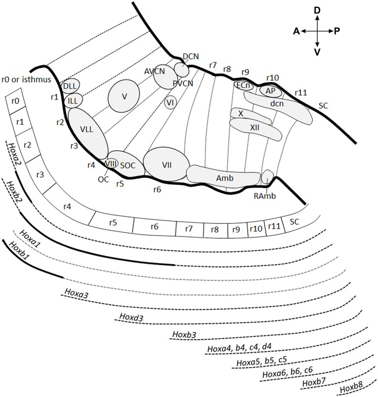
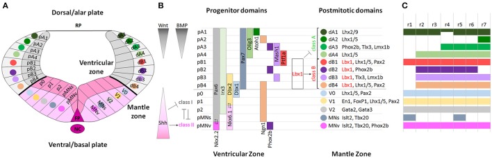
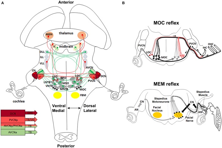
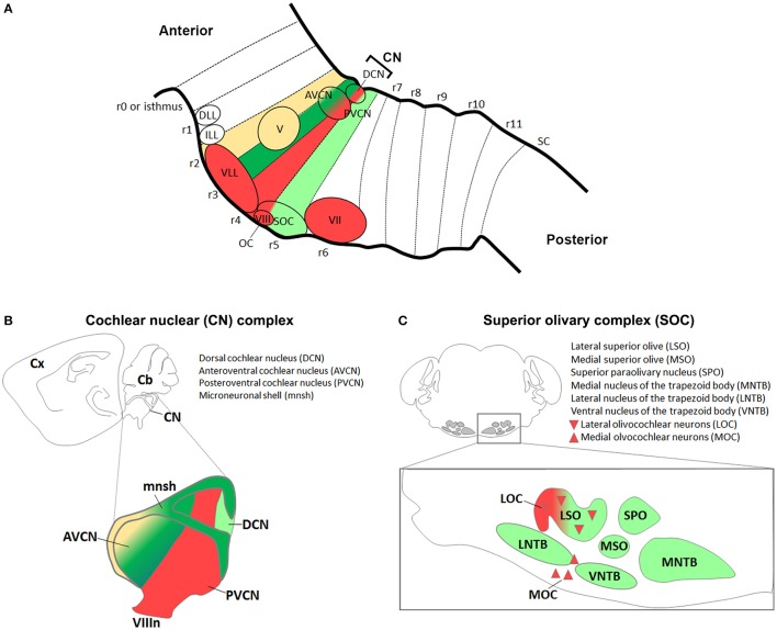
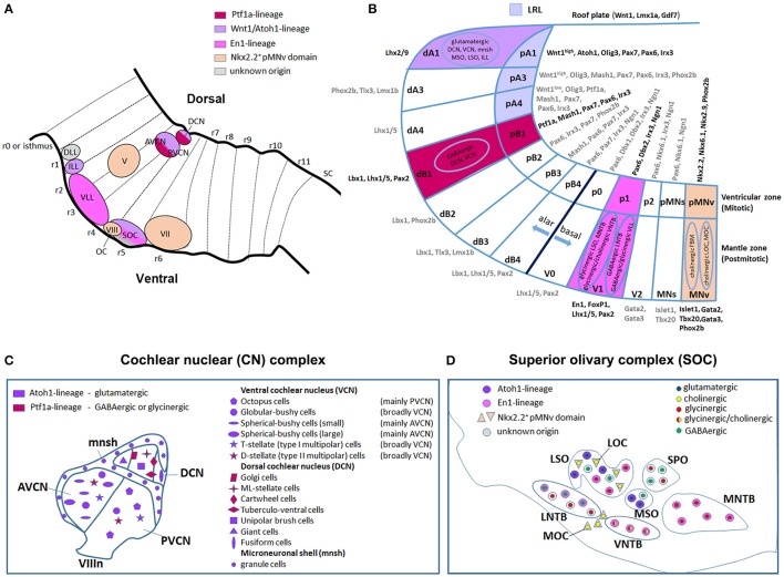
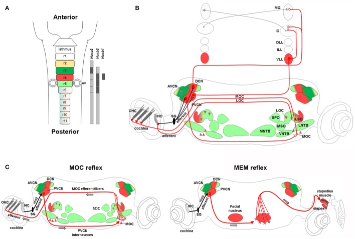
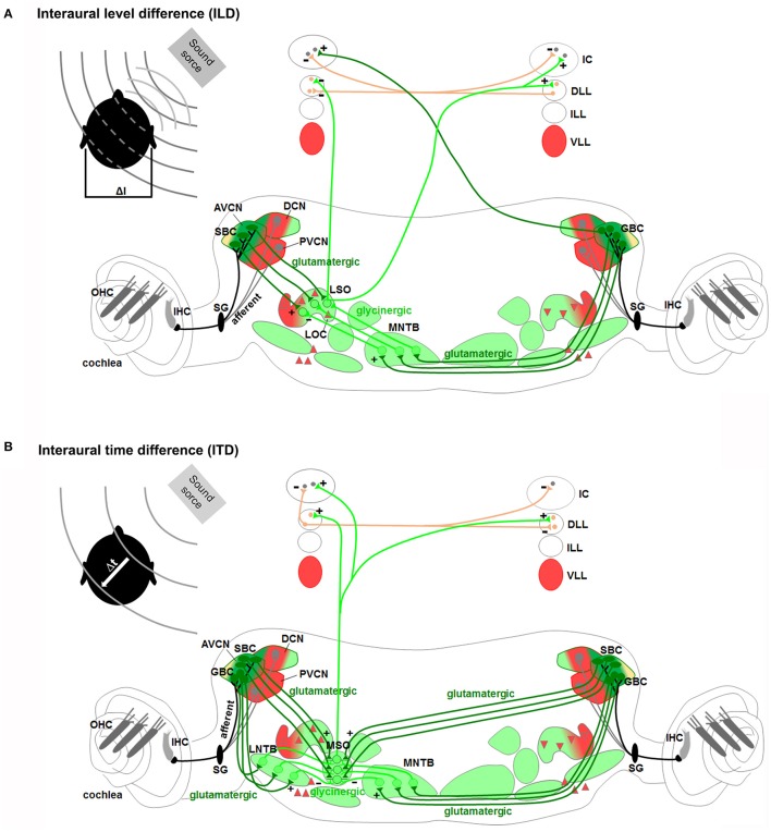
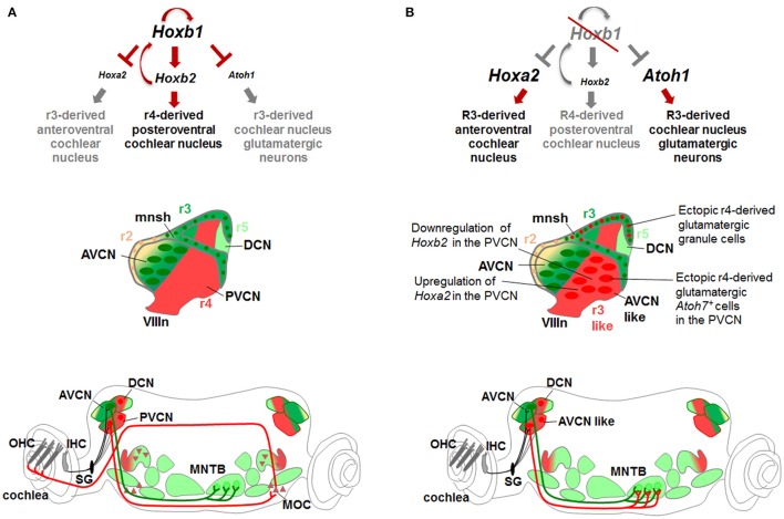
During development, the organization of the auditory system into distinct functional subcircuits depends on the spatially and temporally ordered sequence of neuronal specification, differentiation, migration and connectivity. Regional patterning along the antero-posterior axis and neuronal subtype specification along the dorso-ventral axis intersect to determine proper neuronal fate and assembly of rhombomere-specific auditory subcircuits. By taking advantage of the increasing number of transgenic mouse lines, recent studies have expanded the knowledge of developmental mechanisms involved in the formation and refinement of the auditory system. Here, we summarize several findings dealing with the molecular and cellular mechanisms that underlie the assembly of central auditory subcircuits during mouse development, focusing primarily on the rhombomeric and dorso-ventral origin of auditory nuclei and their associated molecular genetic pathways.

Keywords: DV molecular determinants; Hox genes; central auditory system; mouse lineage tracing; rhombomeres.


Figures

Figure 1
Figure 1
Antero-posterior anatomical subdivision of the pons and medulla oblongata and corresponding Hox gene expression. Schema showing a sagittal view of the hindbrain subdivided into 12 transverse segments: isthmus and r1 (r0–r1) territories, 5 overt rhombomeres (r2–r6), and 5 crypto-rhombomeres (r7–r11) along the AP axis (above). Below, corresponding Hox expression profile correlated to the intersegmentary boundaries and transverse limits of nuclei or intranuclear division. Gray dotted lines refer to Hoxa1 and Hoxb1 downregulation during early development. Thick black lines indicate upregulated expression of Hoxa2 in r3, Hoxb1 in r4 until E12.5 and Hoxb2 in r4-r6. Modified from Tomas-Roca et al. (2016).
Figure 2
Figure 2
Molecular specification of dorso-ventral neuronal cell fate in the hindbrain. (A) Schematic diagram showing the DV organization of a rhombomere from dorsal to ventral: roof plate (RP), dorsal (alar) and ventral (basal) subdomains, floor plate (FP) and notochord (NC). Each domain is formed by progenitors in the ventricular zone and by postmitotic neurons in the mantle zone. (B) Two opposite morphogen gradients of Wnt and BMP (dorsalizing signals) produced by the overlying ectoderm and roof plate, and Shh (ventralizing signal) produced by the notochord and floor plate control the expression of bHLH and HD proteins along the DV axis establishing distinct progenitor domains and directing the differentiation of neuronal subtypes. In the alar plate, dA1-dB4 classes of dorsal neurons are subdivided into class A and class B neurons, respectively repressed or activated by Lbx1 (in red). In the basal plate, HD class I genes are repressed and HD class II genes are activated by Shh signaling, and cross-repressive interactions between class I and class II proteins refine and maintain progenitor domains and determine interneuron (V0-V2), somatic (MNs), and visceral (MNv) motor neuron differentiation. Modified from Takahashi and Osumi (2002), Melton et al. (2004), and Storm et al. (2009). (C) Rhombomeric mapping of hindbrain DV domains, modified from Gray (2008). The DV domains extend differently across rhombomeres along the AP axis.
Figure 3
Figure 3
Anatomical and functional organization of auditory sensorimotor subcircuits. (A) Anatomical overview of auditory pathways described in the text. Monaural (in red) and binaural (in green) systems receive sound information form one or both ears, respectively. Bottom left, diagram showing three major fiber tracts leaving different parts of the CN: trapezoid body (TB), intermediate acoustic stria (IAS), and dorsal acoustic stria (DAS) modified from Cant and Benson (2003). (B) Schema of MOC and MEM reflexes modified from Liberman and Guinan (1998). MOC (contralateral axons in black, and ipsilateral axons in gray) and LOC (red lines) efferent innervation to the cochlea. In the MOC reflex, afferent neurons of the auditory nerve synapse onto cochlear hair cells and project axons centrally to the PVCN. PVCN neurons send axons across the midline to innervate MOC neurons (stars), which mainly project to OHC of the contralateral cochlea and inhibit cochlear responses. The MEM reflex (stapedial reflex pathway) consists of FBM neurons, which are stimulated by VCN interneurons (arrows) and stiffen the ossicular chains by activating the stapedius muscle, thus reducing the sound transmission through the middle ear and offering protection from acoustic overstimulation. AVCNa, anteriorior part of the AVCN; AVCNp, posterior part of the AVCN; PVCNa, anterior part of PVCN; PVCNp, posterior part of PVCN.
Figure 4
Figure 4
Antero-posterior origin of auditory nuclei. (A) Schema showing a sagittal view of the hindbrain in which the color code represents rhombomeric origin of r2- (yellow), r3- (dark green), r4- (red), r5- (light green) derived territories and auditory nuclei. (B,C) Schematic representations of the (B) cochlear nuclear complex (AVCN, PVCN, DCN, and microneuronal shell), and (C) superior olivary complex (LSO, MSO, MNTB, VNTB, LNTB, SPO) and olivocochlear (LOC and MOC) neurons. Colors refer to their rhombomeric origin as described in (A).
Figure 5
Figure 5
Dorso-ventral origin of auditory nuclei. (A) Schema showing a sagittal view of the hindbrain correlating the different auditory nuclei to their DV domains of origin (B) by color code. (B) Schematic transverse section through the developing hindbrain at r4–r6 levels, showing the DV domain of progenitors and derived neurons and their contribution to auditory nuclei, modified from Nothwang (2016). Note that the dA3 domain is absent in r1–r3, while dA4, dB2, dB3, and dB4 are missing in r1 (Sieber et al., ; Gray, 2008). The pMNs domain, dorsal to the pMNv, is only present in r1 and r5 (Takahashi and Osumi, ; Guthrie, 2007). (C,D) Schematic representation of the DV origin and neurotransmitter phenotype of auditory neuronal populations: (C) cochlear nuclear complex [AVCN, PVCN, DCN and microneuronal shell (mnsh)], (D) superior olivary complex (LSO, MSO, MNTB, VNTB, LNTB, SPO) and olivocochlear (LOC and MOC) neurons, modified from Fujiyama et al. (2009) and Altieri et al. (2015), respectively. The same color code is maintained from (A–D).
Figure 6
Figure 6
Rhombomere 4-derived auditory subcircuits. (A) Schema of hindbrain, in which rhombomeres 2–5 are color-coded, and associated Hox genes expression. (B,C) R4-derived subcircuits involved in the transmission of auditory sounds (CN, VLL), protection from acoustic injury (MOC/MEM reflex and LOC), amplification of low-level sounds (MOC innervation to OHC). (C) R4-derived PVCN neurons and motor MOC and FBM neurons contribute to the efferent reflex of MOC and MEM reflex, respectively. Modified from Di Bonito et al. (2013a). Hoxb1 and Hoxb2 act primarily upon assembly of r4-derived structures, contributing to the main pathway of sound transmission, as well as in the establishment of sensorimotor reflex circuits important for cochlea protection and amplification processes.
Figure 7
Figure 7
Rhombomeres 2, 3, and 5-derived auditory subcircuits. The color code of the auditory nuclei is referred to the schema in Figure 6A. (A,B) r2/r3/r5-derived subcircuits involved in the sound localization by (A) interaural level difference (ILD) and (B) interaural time difference (ITD), modified from Grothe et al. (2010). ILD (A) and ITD (B) are processed by neurons of r2/r3-derived AVCN, and r5-derived MNTB and LSO or MSO, respectively. Hoxa2 contributes to AVCN development and connectivity in the sound localization circuit, mainly formed by r2, r3, and r5.
Figure 8
Figure 8
Hox gene networks involved in the assembly of central auditory subcircuits. (A) In r4, Hoxb1 activates Hoxb2 and represses Hoxa2 and Atoh1 preventing the formation of Atoh1-derived glutamatergic neurons normally produced in r3. (B) In the absence of Hoxb1, Atoh1 and Hoxa2 are upregulated while Hoxb2 is downregulated at similar levels than in r3. Ectopic r4-derived granule cells contribute to the microneuronal shell. The r4-derived PVCN acquires “AVCN-like” identity producing ectopic glutamatergic Atoh7+ spherical bushy cells and projecting to the MNTB, a physiologically target of r3-derived AVCN neurons. Modified from Di Bonito et al. (2013a).

Similar articles

Cited by

References

    1. Alasti F., Sadeghi A., Sanati M. H., Farhadi M., Stollar E., Somers T., et al. . (2008). A mutation in HOXA2 is responsible for autosomal-recessive microtia in an Iranian family. Am. J. Hum. Genet. 82, 982–991. 10.1016/j.ajhg.2008.02.015 - DOI - PMC - PubMed
    1. Alaynick W. A., Jessell T. M., Pfaff S. L. (2011). SnapShot: spinal cord development. Cell 146, 178–178 e171. 10.1016/j.cell.2011.06.038 - DOI - PMC - PubMed
    1. Altieri S. C., Jalabi W., Zhao T., Romito-DiGiacomo R. R., Maricich S. M. (2015). En1 directs superior olivary complex neuron positioning, survival, and expression of FoxP1. Dev. Biol. 408, 99–108. 10.1016/j.ydbio.2015.10.008 - DOI - PMC - PubMed
    1. Altieri S. C., Zhao T., Jalabi W., Maricich S. M. (2014). Development of glycinergic innervation to the murine LSO and SPN in the presence and absence of the MNTB. Front. Neural Circuits 8:109. 10.3389/fncir.2014.00109 - DOI - PMC - PubMed
    1. Altieri S. C., Zhao T., Jalabi W., Romito-DiGiacomo R. R., Maricich S. M. (2016). En1 is necessary for survival of neurons in the ventral nuclei of the lateral lemniscus. Dev. Neurobiol. 76, 1266–1274. 10.1002/dneu.22388 - DOI - PMC - PubMed

Publication types

LinkOut - more resources