. 2017 Apr 19:8:669.
doi: 10.3389/fmicb.2017.00669. eCollection 2017.

Fis Regulates Type III Secretion System by Influencing the Transcription of exsA in Pseudomonas aeruginosa Strain PA14

Affiliations

Fis Regulates Type III Secretion System by Influencing the Transcription of exsA in Pseudomonas aeruginosa Strain PA14

Xuan Deng et al. Front Microbiol. .

Abstract

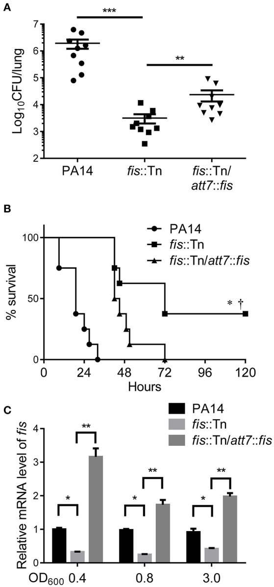
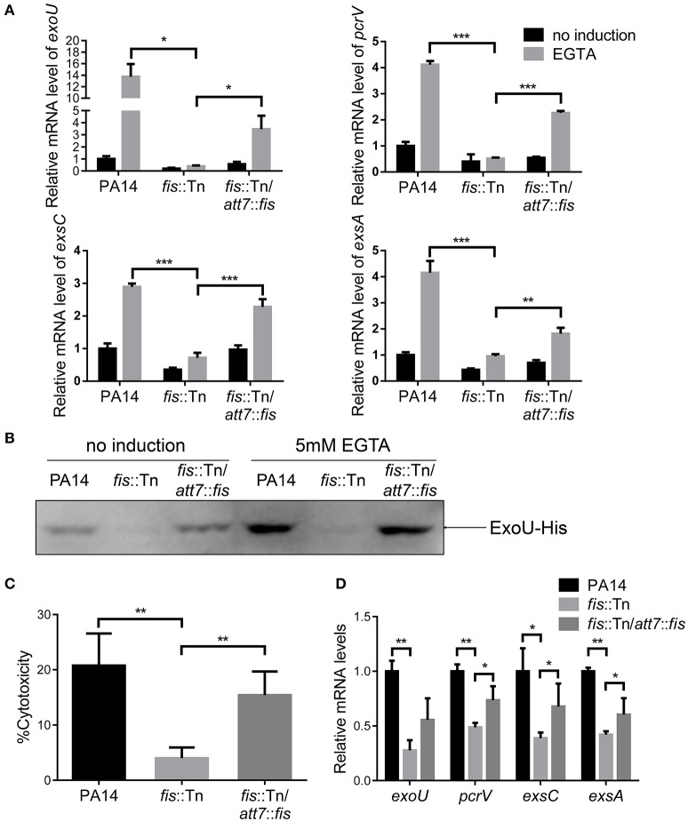
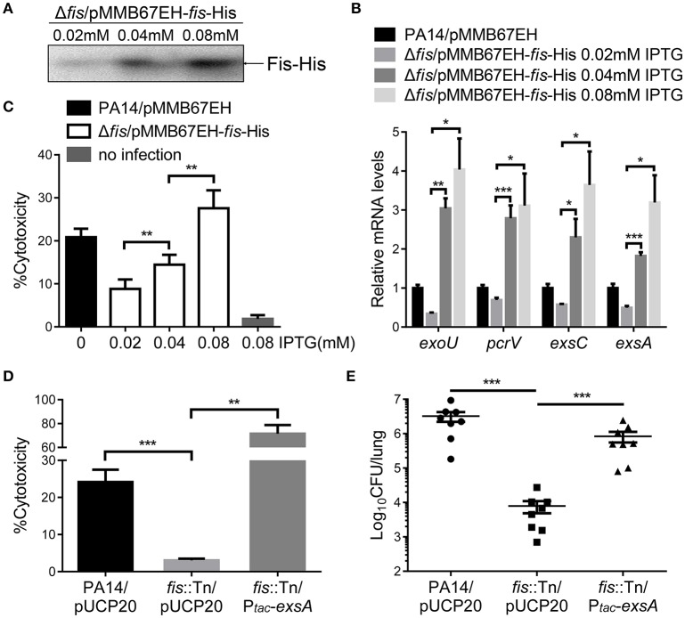
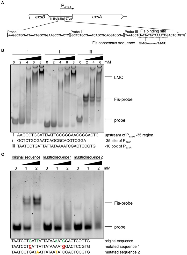
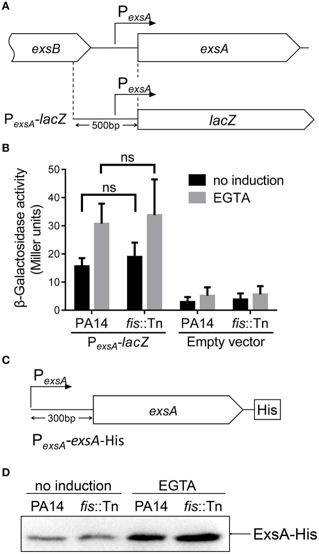
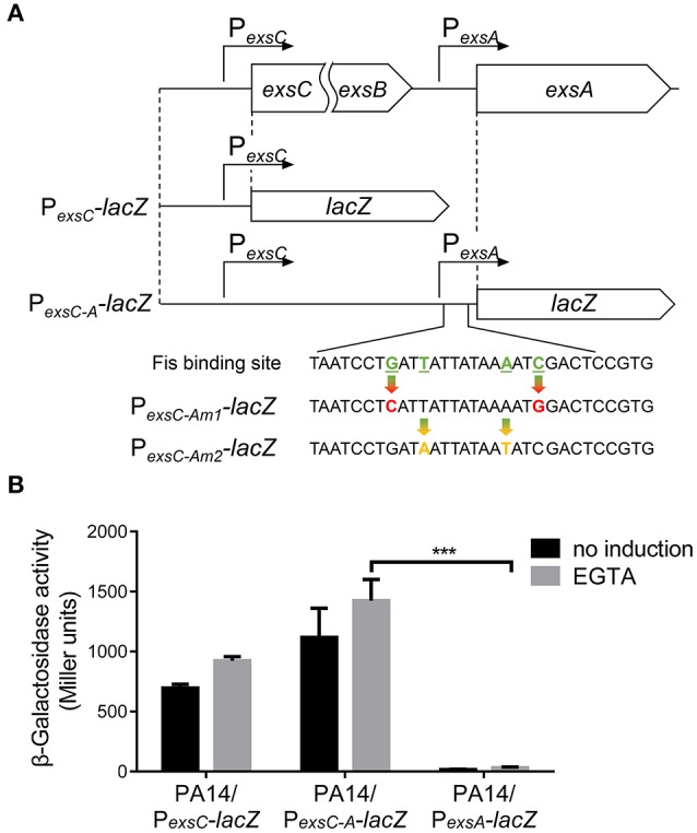
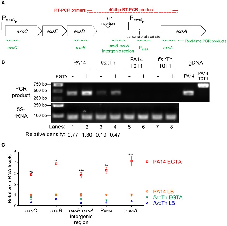
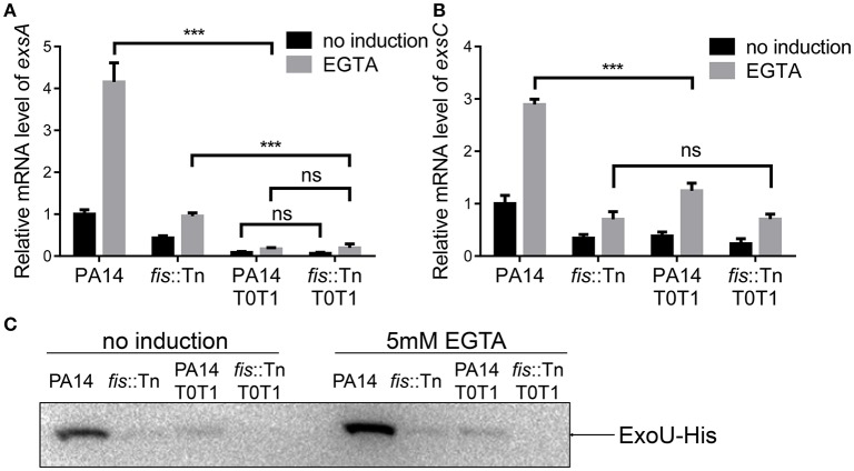
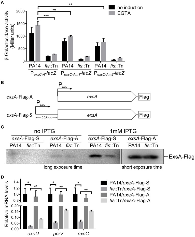
Fis is a versatile DNA binding protein in bacteria. It has been demonstrated in multiple bacteria that Fis plays crucial roles in regulating bacterial virulence factors and optimizing bacterial adaptation to various environments. However, the role of Fis in Pseudomonas aeruginosa virulence as well as gene regulation remains largely unknown. Here, we found that Fis was required for the virulence of P. aeruginosa in a murine acute pneumonia model. Transcriptome analysis revealed that expression of T3SS genes, including master regulator ExsA, was defective in a fis::Tn mutant. We further demonstrate that the continuous transcription of exsC, exsE, exsB, and exsA driven by the exsC promoter was required for the activation of T3SS. Fis was found to specifically bind to the exsB-exsA intergenic region and plays an essential role in the transcription elongation from exsB to exsA. Therefore, we found a novel role of Fis in the regulation of exsA expression.

Keywords: Fis; Pseudomonas aeruginosa; bacterial virulence; exsA transcription; type III secretion system.


Figures

Figure 1
Figure 1
Fis is essential for the bacterial virulence in vivo. (A) Mice were inoculated intranasally with 1 × 10⁷ CFU bacteria of indicated strains. 14 hpi, mice were sacrificed and lungs were isolated and homogenized. Bacterial loads were determined by serial dilution and plating. The central bar indicates the mean, and error bars indicate standard error of the mean. ***p < 0.001; **p < 0.01 by the Mann-Whitney test. (B) Mice were inoculated intranasally with 2 × 10⁷ CFU bacteria of indicated strains. The mice were monitored for 5 days after the infection. *p < 0.001, compared to wild type PA14 by log-rank (Mantel-Cox) test; p < 0.05, compared to the complemented strain (fis::Tn/att7::fis) by log-rank (Mantel-Cox) test. (C) Total RNA was isolated from bacterial culture at indicated optical densities (OD600). cDNA was synthesized with fis and rpsL specific primers. Relative mRNA levels of fis were determined by quantitative real-time PCR. The 30S ribosomal protein coding gene rpsL was used as an internal control. Data represents the mean ± standard deviation from three samples. *p < 0.05; **p < 0.01 by Student's t-test.
Figure 2
Figure 2
Fis is required for T3SS gene expression and bacterial cytotoxicity. (A) Relative mRNA levels of T3SS genes exoU, pcrV, exsC, exsA. Total RNA was isolated from bacteria grown with or without 5 mM EGTA and relative mRNA levels of these genes were determined by quantitative real-time PCR. Data represents the mean ± standard deviation from three samples. *p < 0.05; **p < 0.01; ***p < 0.001 by Student's t-test. (B) PA14, fis::Tn mutant and fis::Tn/att7::fis carrying an exoU-His driven by its native promoter (PexoU-exoU-His) were grown at 37°C with or without 5 mM EGTA for 3 h. Proteins samples from equal amounts of protein were separated by SDS-PAGE and the ExoU-His levels were determined by western blotting analysis using an anti-His antibody. (C) Bacterial cytotoxicity on HeLa cells. HeLa cells were infected with indicated strains at a MOI of 50 for 3 h. The bacterial cytotoxicity was determined by the LDH release assay. Error bars indicate standard deviations of triplicate assays. **p < 0.01 by Student's t-test. (D) Relative mRNA levels of T3SS genes during lung infection. Mice were infected intranasally with indicated strains. 6 hpi, bacteria from BALF were collected, followed by RNA isolation. Relative mRNA levels of exoU, pcrV, exsC, and exsA were determined by quantitative real-time PCR. Data represents the mean ± standard deviation from three independent experiments. *p < 0.05; **p < 0.01 by Student's t-test.
Figure 3
Figure 3
Fis is required for the activation of the T3SS. (A) The strain Δfis/pMMB67EH-fis-His was grown at 37°C with indicated concentrations of IPTG to an OD600 of 1.0. Proteins samples from equal amounts of protein were separated by SDS-PAGE and the Fis-His levels were determined by western blotting analysis using an anti-His antibody. (B) Relative mRNA levels of T3SS genes. Total RNA of indicated strains was isolated from bacteria grown with indicated concentrations of IPTG and mRNA levels of T3SS genes were determined by quantitative real time PCR. Data represents the mean ± standard deviation from three samples. *p < 0.05; **p < 0.01; ***p < 0.001 by Student's t-test. (C) Bacterial cytotoxicity on HeLa cells. HeLa cells were infected with indicated strains with indicated concentration of IPTG at a MOI of 50 for 3 h. The bacterial cytotoxicity was determined by the LDH release assay. Error bars indicate standard deviations of triplicate assays. **p < 0.01, by Student's t-test. (D) HeLa cells were infected with indicated strains at a MOI of 50 for 3 h. The bacterial cytotoxicity was determined by the LDH release assay. Error bars indicate standard deviations of triplicate assays. **p < 0.01; ***p < 0.001 by Student's t-test. (E) Mice were inoculated intranasally with 1 × 10⁷ CFU bacteria of indicated strains. 14 hpi, mice were sacrificed and lungs were isolated and homogenized. Bacterial loads were determined by serial dilution and plating. The central bar indicates the mean, and error bars indicate standard error of the mean. ***p < 0.001 by the Mann-Whitney test.
Figure 4
Figure 4
Fis directly interacts with exsA promoter region. (A) Diagram of the exsA promoter region. PexsA is indicated by an arrow. Sequences of the probes used in EMSA and the consensus Fis binding sequence are shown. The transcription start site of exsA is indicated by an asterisk. Fragment i, ii, and iii represent DNA probes used in the EMSA. (B) Fis was incubated with probe i, ii, or iii for 30 min at 25°C. Arrows indicate the positions of unbound probes or the Fis-probe complex and LMC. (C) Binding of the Fis to the DNA fragment containing PexsA -10 box original and mutated sequences. Colored underlined letters represent mutated nucleotides. Arrows indicate positions of the Fis-probe complex and unbound probes, respectively.
Figure 5
Figure 5
Role of Fis in the regulation of PexsA. (A) Diagram of the PexsA-lacZ transcriptional fusion. (B) PA14 and the fis::Tn mutant carrying the PexsA-lacZ fusion reporter or empty vector (promoterless lacZ) were grown at 37°C with or without 5 mM EGTA for 3 h. The values (Miller units) are the means of three experiments. ns, not significant by Student's t-test. (C) Diagram of the PexsA-exsA-His construct. The exsA open reading frame with its upstream 300 bp region was fused with a 6 × His tag at the C-terminus. (D) PA14 and the fis::Tn mutant carrying an exsA-His driven by its native promoter (PexsA-exsA-His) were grown at 37°C with or without 5 mM EGTA for 3 h. Proteins samples from equal amounts of protein were separated by SDS-PAGE. The ExsA-His levels were determined by western blotting analysis with an anti-His antibody.
Figure 6
Figure 6
Transcription driven by the exsC or exsA promoter. (A) Diagram of the PexsC-lacZ and PexsC-A-lacZ transcriptional fusions. Point mutations of Fis binding site in PexsC-Am1-lacZ and PexsC-Am2-lacZ transcriptional fusions are indicated by arrows. (B) PA14 carrying PexsC-lacZ, PexsC-A-lacZ or PexsA-lacZ were grown at 37°C with or without 5 mM EGTA and assayed for β-galactosidase activities. The reported values (Miller units) are the means of results of at least three independent experiments. ***p < 0.001 by Student's t-test.
Figure 7
Figure 7
Transcription of exsC, exsE, exsB and exsA. (A) Diagram of the exsCEBA operons. PexsC and PexsA are indicated by arrows. Red arrows indicate the directions and locations of the primers for RT-PCR. Insertion site of the transcriptional terminator T0T1 is indicated. Green wavy lines indicate positions of real time PCR products. (B) Total RNA was isolated from indicated strains grown with or without 5 mM EGTA for 3 h. cDNA was synthesized and used as templates in PCR. The 5S rRNA was used as an internal control. The density of each band in lanes 1–4 was determined with ImageJ (ImageJ software k1.45). The relative density was calculated by dividing the density of each exsB-exsA RT-PCR product by that of the corresponding 5S rRNA RT-PCR product (C) Relative mRNA levels of five regions within the exsCEBA operon. PA14 and fis::Tn mutant were grown in the presence or absence of 5 mM EGTA. The value of each tested fragment represents the RNA level relative to that in wild type PA14 grown in LB medium. Data represents the mean ± standard deviation from three independent experiments. **p < 0.01, ***p < 0.001, compared to wild type PA14 grown in LB medium by Student's t-test.
Figure 8
Figure 8
exsA transcription relies mainly on PexsC. (A) Relative mRNA levels of exsA. Total RNA of indicated strains was isolated from bacteria grown with or without 5 mM EGTA and mRNA levels of exsA were determined by quantitative real time PCR. Data represents the mean ± standard deviation from three samples. ***p < 0.001 by Student's t-test. (B) Relative mRNA levels of exsC. Total RNA of indicated strains was isolated from bacteria grown with or without 5 mM EGTA and mRNA levels of exsC were determined by quantitative real time PCR. Data represents the mean ± standard deviation from three samples. ***p < 0.001 by Student's t-test. (C) PA14, fis::Tn mutant, PA14 T0T1 or fis::Tn T0T1 carrying an exoU-His driven by its native promoter (PexoU-exoU-His) were grown at 37°C with or without 5 mM EGTA for 3 h. Proteins samples from equal amounts of protein were separated by SDS-PAGE. ExoU-His levels were determined by western blotting analysis using an anti-His antibody.
Figure 9
Figure 9
Role of Fis in the transcription elongation from exsB to exsA. (A) PA14 and fis::Tn mutant carrying PexsC-A-lacZ, PexsC-Am1-lacZ, or PexsC-Am2-lacZ transcriptional reporters were grown at 37°C with or without 5 mM EGTA. The values are the means of at least three independent experiments. ***p < 0.001; **p < 0.01 by Student's t-test. (B) Constructs of exsA-Flag-S and exsA-Flag-A. exsA-Flag-S contains exsA ORF and 225 bp upstream fragment fused with tac promoter and exsA-Flag-A contains exsA ORF only fused with tac promoter. (C) PA14 and fis::Tn mutant carrying plasmids exsA-Flag-S or exsA-Flag-A were grown with or without 1 mM IPTG for 3 h. ExsA-Flag levels were determined by western blot using an anti-Flag antibody. The amounts of protein in different samples were equal. (D) Relative mRNA levels of T3SS genes exoU, pcrV, exsC, exsA. Total RNA was isolated from bacteria grown with 1 mM IPTG and relative mRNA levels of these genes were determined by quantitative real-time PCR. Data represents the mean ± standard deviation from three samples. *p < 0.05, **p < 0.01 by Student's t-test.

Similar articles

Cited by

References

    1. Agnello M., Finkel S. E., Wong-Beringer A. (2016). Fitness cost of fluoroquinolone resistance in clinical isolates of Pseudomonas aeruginosa differs by Type III secretion genotype. Front. Microbiol. 7:1591. 10.3389/fmicb.2016.01591 - DOI - PMC - PubMed
    1. Aiyar S. E., McLeod S. M., Ross W., Hirvonen C. A., Thomas M. S., Johnson R. C., et al. . (2002). Architecture of Fis-activated transcription complexes at the Escherichia coli rrnB P1 and rrnE P1 promoters. J. Mol. Biol. 316, 501–516. 10.1006/jmbi.2001.5390 - DOI - PubMed
    1. Anantharajah A., Mingeot-Leclercq M. P., Van Bambeke F. (2016). Targeting the type three secretion system in Pseudomonas aeruginosa. Trends Pharmacol. Sci. 37, 734–749. 10.1016/j.tips.2016.05.011 - DOI - PubMed
    1. Anderson G. G., Yahr T. L., Lovewell R. R., O'Toole G. A. (2010). The Pseudomonas aeruginosa magnesium transporter MgtE inhibits transcription of the type III secretion system. Infect. Immun. 78, 1239–1249. 10.1128/IAI.00865-09 - DOI - PMC - PubMed
    1. Audic S., Claverie J. M. (1997). The significance of digital gene expression profiles. Genome Res. 7, 986–995. - PubMed

LinkOut - more resources