Herlyn-Werner-Wunderlich Syndrome: Sonographic and Magnetic Resonance (MR) Imaging Findings of This Rare Urogenital Anomaly
- PMID: 28469738
- PMCID: PMC5402867
- DOI: 10.12659/PJR.899889
Herlyn-Werner-Wunderlich Syndrome: Sonographic and Magnetic Resonance (MR) Imaging Findings of This Rare Urogenital Anomaly
Abstract
Background: Herlyn-Werner-Wunderlich syndrome is a rare congenital urogenital anomaly characterised by uterus didelphys with blind hemivagina and ipsilateral renal agenesis. Children usually have progressive pelvic pain after menarche, palpable mass due to hemihaemato(metro)colpos or pelvic inflammatory disease. The diagnosis usually requires a suspicion of this rare genitourinary syndrome.
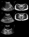
Case reports: We present ultrasonography and MR imaging findings of this rare anomaly in two cases.
Conclusions: Early recognition of this rare syndrome can lead to an immediate, proper surgical intervention and is necessary to prevent complications and preserve future fertility. Ultrasound and MR imaging findings can collectively delineate uterine morphology, indicate the absence of ipsilateral kidney and show obstructed hemivagina.
Keywords: Magnetic Resonance Imaging; Ultrasonography, Doppler, Color; Urogenital Abnormalities.
Conflict of interest statement
Statement No funding was received from any source. The authors declare no conflicts of interest.
Figures


References
-
- Sadler TW, Langman J, editors. Langman’s medical embryology. 8th edn. Lippincott Williams & Wilkins; Philadelphia: 2000.
-
- Kiechl-Kohlendorfer U, Geley TE, Unsinn KM, et al. Diagnosing neonatal female genital anomalies using saline enhanced sonography. Am J Roentgenol. 2001;177:1041–44. - PubMed
-
- Purslow CE. A case of unilateral haematocolpos, haematometra and haematosalpinx. J Obstet Gyaecol Br Emp. 1922;29:643.
-
- Ballesio L, Andreoli C, De Cicco ML, et al. Hematocolpos in double vagina associated with uterus didelphus: US and MR findings. Eur J Radiol. 2003;45:150–53. - PubMed
Publication types
LinkOut - more resources
Full Text Sources
Other Literature Sources
