. 2017 May 2:8:8.
doi: 10.1186/s13227-017-0071-0. eCollection 2017.

Developing an ancient epithelial appendage: FGF signalling regulates early tail denticle formation in sharks

Affiliations

Developing an ancient epithelial appendage: FGF signalling regulates early tail denticle formation in sharks

Rory L Cooper et al. Evodevo. .

Abstract

Background: Vertebrate epithelial appendages constitute a diverse group of organs that includes integumentary structures such as reptilian scales, avian feathers and mammalian hair. Recent studies have provided new evidence for the homology of integumentary organ development throughout amniotes, despite their disparate final morphologies. These structures develop from conserved molecular signalling centres, known as epithelial placodes. It is not yet certain whether this homology extends beyond the integumentary organs of amniotes, as there is a lack of knowledge regarding their development in basal vertebrates. As the ancient sister lineage of bony vertebrates, extant chondrichthyans are well suited to testing the phylogenetic depth of this homology. Elasmobranchs (sharks, skates and rays) possess hard, mineralised epithelial appendages called odontodes, which include teeth and dermal denticles (placoid scales). Odontodes constitute some of the oldest known vertebrate integumentary appendages, predating the origin of gnathostomes. Here, we used an emerging model shark (Scyliorhinus canicula) to test the hypothesis that denticles are homologous to other placode-derived amniote integumentary organs. To examine the conservation of putative gene regulatory network (GRN) member function, we undertook small molecule inhibition of fibroblast growth factor (FGF) signalling during caudal denticle formation.

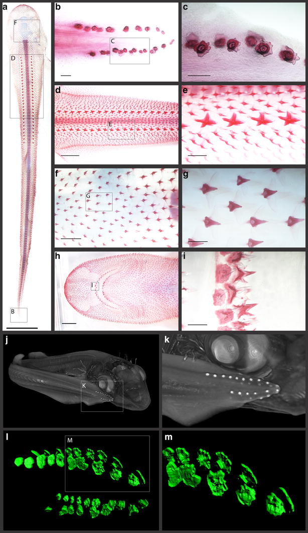
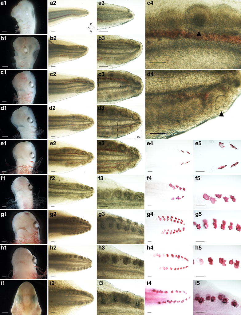
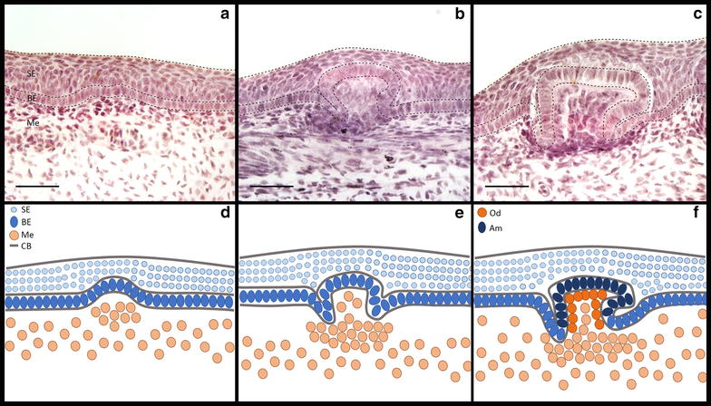
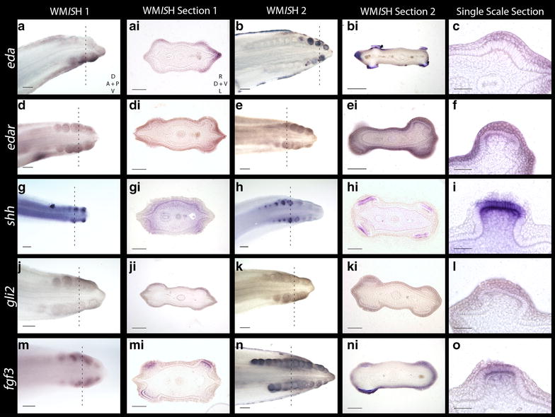
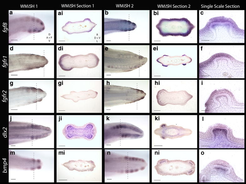
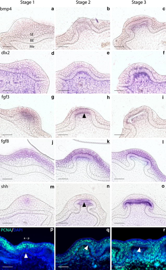
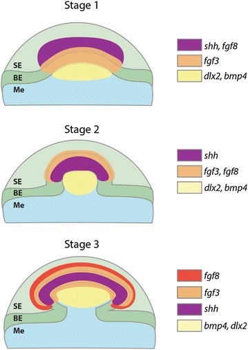
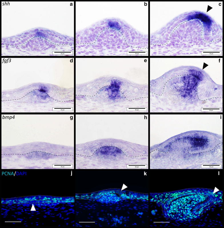
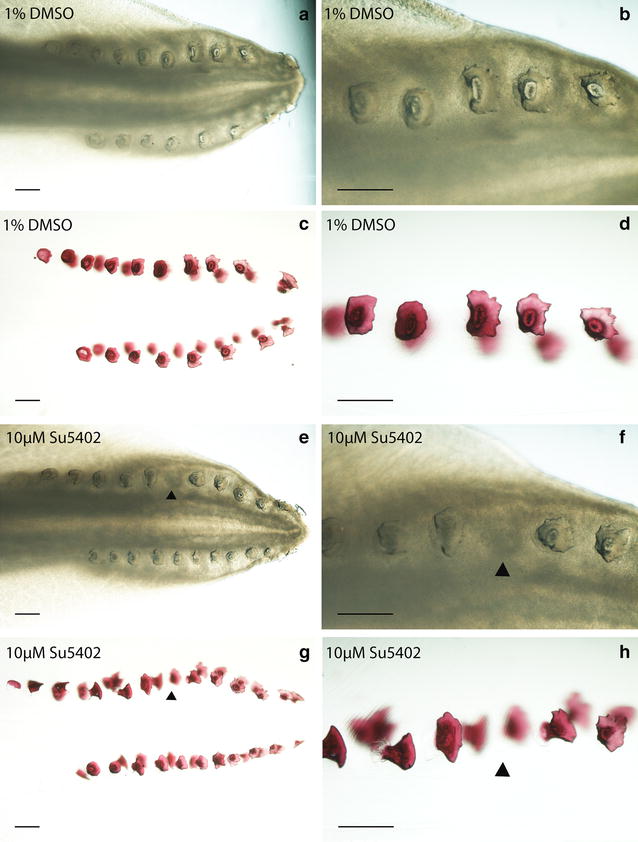
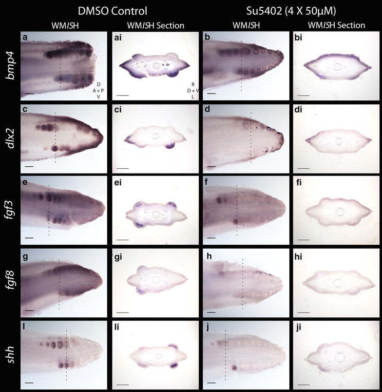
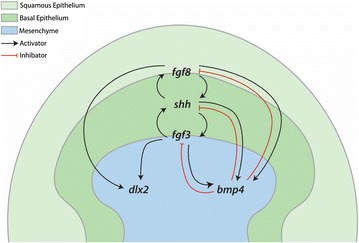
Results: We show that during early caudal denticle morphogenesis, the shark expresses homologues of conserved developmental gene families, known to comprise a core GRN for early placode morphogenesis in amniotes. This includes conserved expression of FGFs, sonic hedgehog (shh) and bone morphogenetic protein 4 (bmp4). Additionally, we reveal that denticle placodes possess columnar epithelial cells with a reduced rate of proliferation, a conserved characteristic of amniote skin appendage development. Small molecule inhibition of FGF signalling revealed placode development is FGF dependent, and inhibiting FGF activity resulted in downregulation of shh and bmp4 expression, consistent with the expectation from comparison to the amniote integumentary appendage GRN.

Conclusion: Overall, these findings suggest the core GRN for building vertebrate integumentary epithelial appendages has been highly conserved over 450 million years. This provides evidence for the continuous, historical homology of epithelial appendage placodes throughout jawed vertebrates, from sharks to mammals. Epithelial placodes constitute the shared foundation upon which diverse vertebrate integumentary organs have evolved.

Keywords: Anatomical placode; Dermal denticle; Epithelial appendage; Homology; Shark.


Figures

Fig. 1
Fig. 1
Odontode diversity of the pre-hatchling Catshark (S. canicula). Samples ai are cleared and stained for calcium-rich tissue using alizarin red dye. Samples jk are computerised tomography (CT) scans of a Stage 32 whole embryo, and samples lm are light sheet fluorescence microscopy (LSFM) images of caudal denticles of a Stage 31 embryo, stained with alizarin red. The pre-hatchling (a) possesses three major external denticle types. The caudal denticles are the first to emerge, appearing on either side of the tip of the tail in dorsal and ventral rows (b, c, jm) [23]. These denticles are not strongly polarised, although cusps generally point towards the posterior [27]. Next, the dorsal denticles emerge along the trunk of the embryo in two polarised rows (d, e). Finally, general body denticles emerge just before hatching, covering the whole body (f, g). These denticles are also highly polarised. Teeth emerge in the jaws at a similar stage to general body denticles (h, i). The scale bar for a = 1000 µm, b, c, g and i = 200 µm, d and h = 2500 µm, e and f = 500 µm
Fig. 2
Fig. 2
Sequential development of caudal denticles in the Catshark. As the embryo develops from Stage 27 (a) to Stage 33 (i), the gills proliferate, the eyes are encircled with pigment of increasing darkness and the rostrum protrudes anterior to the mouth [23]. During this period, caudal denticles develop from posterior to anterior in dorsal and ventral rows, on either side of the tail tip. At early Stage 27, no placodes can be detected (a2a2). Epithelial thickenings then form from posterior to anterior (b2b3, c2c4). c4 shows a magnified view of c3, highlighting an individual placode (marked with an arrowhead). These placodes then accumulate their first layers of mineralised tissue during morphogenesis (d2d4). d4 shows a magnified view of d3, highlighting a mineralising placode (marked with an arrowhead). Mineralisation of denticles also occurs sequentially from posterior to anterior (e2e3, f2f3, g2g3, h2h3 and i2i3) and can be highlighted with alizarin red staining for calcium-rich tissue (e4e5, f4f5, g4g5, h4h5 and i4i5). For the axis, D dorsal, V ventral, P posterior and A anterior. Scale bars are 1000 µm for a1, b1, c1, d1, e1, f1, g1, h1 and i1 and 200 µm for all other images
Fig. 3
Fig. 3
Morphogenesis of a caudal denticle. Caudal denticle placodes consist of a squamous epithelium (SE) overlying columnar cells of the basal epithelium (BE), which overlies the mesenchyme (Me) (a, d). During placode morphogenesis, condensing mesenchymal cells aggregate below columnar cells of the basal epithelium epithelial. The basal epithelium undergoes growth and folding (b, e) to form the posterior facing cusp (c, f). CB is cell layer boundary. Ameloblasts (Am) in the basal epithelial cusp (c, f) and odontodes (Od) in the papilla underlying the basal epithelium produce enameloid and dentine, respectively, to mineralise the unit [40]. Scale bars are 50 µm
Fig. 4
Fig. 4
Gene expression analyses of early morphogenesis of caudal denticles. Expression of eda and its receptor edar are observed in the epithelium during early placode morphogenesis (af). eda can also be seen in tissue undergoing mineralisation later in morphogenesis (bbi). shh is first observed in the epithelium during early morphogenesis, before becoming restricted to the basal epithelium later in morphogenesis (gi). gli2 is also seen in the epithelium early during placode formation (jl). Expression of fgf3 is first seen in the epithelium, before moving to both the epithelium and mesenchyme later in placode morphogenesis (mo). The dashed lines show where in the WMISH the section was taken. WMISH Section 1 represents a younger stage specimen than WMISH Section 2. For the WMISH, D dorsal, V ventral, A anterior and P posterior. For WMISH sections, R right, L left, D dorsal and V ventral. For scale bars, a, b, d, e, g, h, j, k, m, n = 200 µm, ai, bi, di, ei, gi, hi, ji, ki, mi, ni = 100 µm, and c, f, i, l, o = 50 µm
Fig. 5
Fig. 5
fgf8 signalling is largely retained in the epithelium throughout (ac). fgfr1 and fgfr2 are both seen in the epithelium during early denticle morphogenesis (di). Expression of dlx2 is restricted to the mesenchyme throughout early placode morphogenesis (jl). Similarly, bmp4 is observed in the mesenchyme during early placode morphogenesis (mo). The dashed lines show where in the WMISH the section was taken. WMISH Section 1 represents a younger stage specimen than WMISH Section 2. For the WMISH, D dorsal, V ventral, A anterior and P posterior. For WMISH sections, R right, L left, D dorsal and V ventral. For scale bars, a, b, d, e, g, h, j, k, m, n = 200 µm, ai, bi, di, ei, gi, hi, ji, ki, mi, ni = 100 µm, and c, f, i, l and o = 50 µm
Fig. 6
Fig. 6
Gene expression/PCNA analysis of early caudal denticle morphogenesis. Gene expression is shown in 30-µm transverse sections of wild-type S. canicula embryo tails post-WMISH, to highlight progressive stages of caudal denticle morphogenesis from the initial epithelial thickening. bmp4 and dlx2 expression is restricted to the mesenchyme throughout morphogenesis (Me) (af). shh and fgf8 are first observed in the squamous epithelium (SE) before becoming restricted to the basal epithelium (BE) (mo, jl). Expression of fgf3 begins in the squamous and basal epithelium and is subsequently observed throughout the epithelium and mesenchyme (gi). PCNA immunofluorescence is observed in the epithelium and mesenchyme throughout morphogenesis (pr). Reduced activity (marked with an arrowhead) was noted in columnar cells of the epithelium during early morphogenesis (p) and in a central region of columnar cells of the basal epithelium during later morphogenesis (qr). This region (q) overlaps with fgf3 and shh expression in the basal epithelium (k, n) (marked with an arrowhead) and may be indicative of a basic primary enameloid knot. a is anterior, and p is posterior. Dashed lines separate the squamous epithelium (SE), basal epithelium (BE) and mesenchyme (Me). All scale bars are 50 µm in length
Fig. 7
Fig. 7
Schematic diagram representing gene expression during early morphogenesis of caudal denticles. This diagram summarises the results from Figs. 4 and 5, representing expression of fgf3, fgf8, shh, bmp4 and dlx2 throughout progressive stages of early morphogenesis. SE is the squamous epithelium, BE is the basal epithelium and Me is the mesenchyme
Fig. 8
Fig. 8
Gene expression analysis of putative placode GRN members, during general body denticle development. Section in situ hybridisation (SISH) was undertaken during early development of body denticles. Expression of shh was epithelial throughout development (ac), whereas fgf3 was observed in both the epithelium and mesenchyme (df). bmp4 was mesenchymal throughout early morphogenesis (gi). PCNA immunoreactivity was observed in the epithelial cells and condensing mesenchyme of emerging denticles (jl). Reduced immunoreactivity was noted in columnar cells of the basal epithelium during placode formation (j) (white arrowed). fgf3 and shh expression marks enameloid knot-like cells of the epithelium associated with denticle morphogenesis (c, f), which also show reduced PCNA immunoreactivity (l), characteristic of this signalling centre (black arrowheads). The dashed line separates the epithelium from the underlying mesenchyme (ai), as well as the basal epithelium and squamous epithelium (jl). All scale bars are 50 µm in length except for image i for which the scale bar is 100 µm
Fig. 9
Fig. 9
Phenotypic effect of FGF inhibition via SU5402 treatment (10 µM) on caudal denticle development. The DMSO control specimen shown after fixation (a, b) and cleared and stained for calcium-rich tissue using alizarin red (c, d) possesses a full sequence of caudal denticles. However, the specimen treated with the FGF antagonist SU5402 has the 6th denticle missing from the sequence, shown after fixation (e, f) and cleared and stained (g, h). This is marked with a black arrowhead. This denticle knockout corresponds to the stage at which treatment occurred, and was observed in 40% of Su5402-treated specimens (n = 5). Scale bars are 200 µm in length
Fig. 10
Fig. 10
Genetic effect of FGF inhibition via SU5402 treatment (4 × 50 µM) on caudal denticle development. There was a reduction in staining intensity of bmp4 (abi), dlx2 (cdi), fgf3 (efi), fgf8 (ghi) and shh (iji) in SU5402-treated specimens compared to DMSO-treated controls. We propose this resulted from the interruptions to the following GRN interactions. SU5402 inhibits FGF activity by blocking FGFR activity, thereby reducing expression of fgf3 and fgf8 (ehi). This reduced shh and dlx2 expression as a FGF—shh positive feedback loops that would normally promote shh and dlx2 expression (as observed during feather development) were interrupted (iji, cdi) [78]. The fgf4shh positive feedback loop that promotes bmp4 was also interrupted by the SU5402 treatment, reducing shh and bmp4 expression (iji, abi) [36]. SU5402-treated and DMSO control specimens both underwent the colour reaction of the WMISH protocol for the same length of time. The dashed lines show where the section was taken from. Scale bars for WMISH are 200 µm in length, and for the WMISH sections they are 100 µm in length
Fig. 11
Fig. 11
Putative relationship between FGF and associated GRN components during caudal denticle morphogenesis. As observed widely throughout epithelial appendage development, for example during feather placode development, FGF—shh positive feedback loops which promote mesenchymal bmp4 are likely to promote early caudal denticle placode morphogenesis. bmp4 may then act as an internal inhibitor, limiting the size of the final unit. FGF signalling can also promote mesenchymal expression of dlx2. This is a hypothetical GRN based on findings from previous research, gene expression data (Figs. 4, 5, 6, 8) and small molecule inhibition of FGF signalling during early caudal denticle morphogenesis, using SU5402 (Figs. 9, 10)

Similar articles

Cited by

References

    1. Pispa J, Thesleff I. Mechanisms of ectodermal organogenesis. Dev Biol. 2003;262:195–205. doi: 10.1016/S0012-1606(03)00325-7. - DOI - PubMed
    1. Biggs LC, Mikkola ML. Early inductive events in ectodermal appendage morphogenesis. Semin Cell Dev Biol. 2014;25–26:11–21. doi: 10.1016/j.semcdb.2014.01.007. - DOI - PubMed
    1. Milinkovitch MC, Manukyan L, Debry A, Di-Poï N, Martin S, Singh D, et al. Crocodile head scales are not developmental units but emerge from physical cracking. Science. 2013;339:78–81. doi: 10.1126/science.1226265. - DOI - PubMed
    1. Di-Poï N, Milinkovitch MC. The anatomical placode in reptile scale morphogenesis indicates shared ancestry among skin appendages in amniotes. Sci Adv. 2016;2:1–8. doi: 10.1126/sciadv.1600708. - DOI - PMC - PubMed
    1. Sengel P. Pattern formation in skin development. Int J Dev Biol. 1990;34:33–50. - PubMed