

Gastric outlet obstruction secondary to caecal herniation into the lesser sac
- PMID: 28469837
- PMCID: PMC5406613
- DOI: 10.1093/jscr/rjx076
Gastric outlet obstruction secondary to caecal herniation into the lesser sac
Abstract
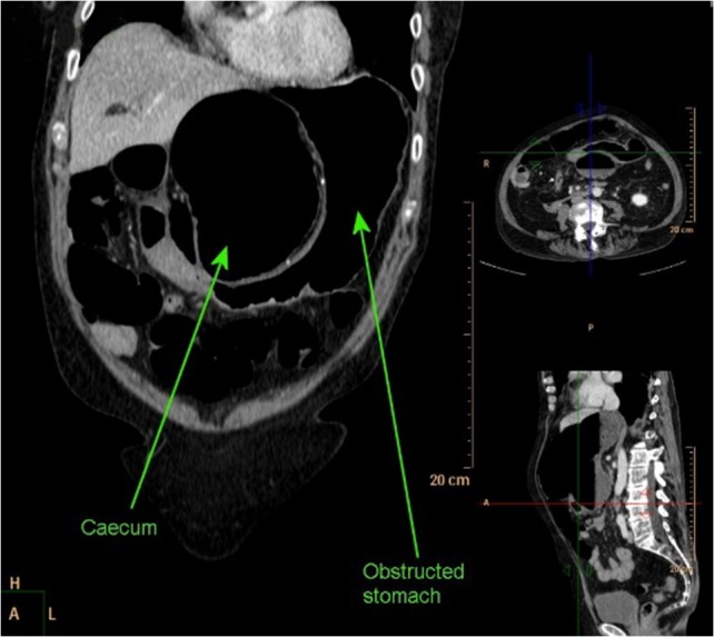
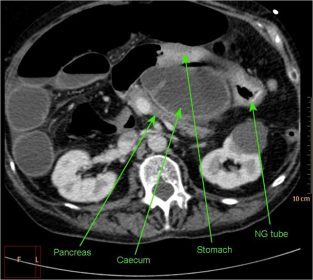
Internal hernias comprise 1% of hernias, 8% of which are through the foramen of Winslow into the lesser sac. These hernias can mimic gastric outlet obstruction and cause associated morbidity. In this case, we describe a caecal herniation into the lesser sac presenting as true gastric outlet obstruction in a 69-year-old female. Initial computed tomography (CT) imaging demonstrated a distended stomach with collapsed small bowel representing likely gastric outlet obstruction. Nasogastric tube insertion decompressed the stomach but the clinical picture progressed to that of small bowel obstruction with generalized abdominal distension and hypoactive bowel sounds. Repeat CT demonstrated caecal herniation into the lesser sac. This was confirmed at exploratory laparotomy with the caecum found in the lesser sac via the foramen of Winslow. The caecum was grossly ischaemic with patchy necrosis. A limited right hemicolectomy was performed. The patient made an uncomplicated recovery and was discharged on the eighth post-operative day.
Figures
Similar articles
-
A case report of caecal herniation through the foramen of Winslow.Ann R Coll Surg Engl. 2018 Jul;100(6):e142-e144. doi: 10.1308/rcsann.2018.0071. Epub 2018 Apr 16. Ann R Coll Surg Engl. 2018. PMID: 29658335 Free PMC article.
-
Herniation of the caecum and ascending colon through the foramen of Winslow: a case report and review.Ann R Coll Surg Engl. 2020 Feb;102(2):e39-e41. doi: 10.1308/rcsann.2019.0123. Epub 2019 Sep 18. Ann R Coll Surg Engl. 2020. PMID: 31532226 Free PMC article. Review.
-
Internal herniation of the cecum through the foramen of Winslow-a case report.J Surg Case Rep. 2021 Oct 31;2021(10):rjab459. doi: 10.1093/jscr/rjab459. eCollection 2021 Oct. J Surg Case Rep. 2021. PMID: 34733471 Free PMC article.
-
Lesser sac herniation: a rare cause of acute abdomen and bowel perforation.BJR Case Rep. 2016 Jun 29;3(1):20150501. doi: 10.1259/bjrcr.20150501. eCollection 2017. BJR Case Rep. 2016. PMID: 30363323 Free PMC article.
-
An adolescent with ileum herniation through foramen of winslow: A case report and literature review.Niger J Clin Pract. 2022 Aug;25(8):1372-1376. doi: 10.4103/njcp.njcp_1778_21. Niger J Clin Pract. 2022. PMID: 35975390 Review.
Cited by
-
Internal Herniation of the Transverse Colon and Stricture of the Gastric Body: A Case Report.Cureus. 2024 Mar 22;16(3):e56712. doi: 10.7759/cureus.56712. eCollection 2024 Mar. Cureus. 2024. PMID: 38646350 Free PMC article.
-
A rare case of a volvulus of the ascending colon strangulated through the foramen of Winslow: "A case report and comprehensive literature review of this rare entity".Radiol Case Rep. 2025 Jun 3;20(9):4144-4151. doi: 10.1016/j.radcr.2025.05.026. eCollection 2025 Sep. Radiol Case Rep. 2025. PMID: 40528904 Free PMC article.
References
-
- Evrard V. Herniation through the foramen of Winslow. Report of two cases. Dis Colon Rectum 1996;39:1055–7. - PubMed
-
- Salar O. Internal hernias: a brief review. Hernia 2013;17:373–7. doi:10.1007/s10029-012-1023-1. - DOI - PubMed
-
- Meyers MA. Dynamic Radiology of the Abdomen: Normal and Pathologic Anatomy. 4th edn New York, NY: Springer, 1994.
-
- Powell-Brett S, Royle J. Caecum herniation through the Foramen of Winslow. J Surg Case Rep 2012;2012:3 pages.10.1093/jscr/rjs01. - DOI - PMC - PubMed
-
- Ray K. Gastric outlet obstruction from a caecal volvulus, herniated through epiploic foramen: a case report. BMJ Case Rep 2009;2009 pii: bcr05.2009.1880. doi: 10.1136/bcr.05.2009.1880. - DOI - PMC - PubMed
Publication types
LinkOut - more resources
Full Text Sources
Other Literature Sources
