Central nervous system involvement of acute promyelocytic leukemia, three case reports
- PMID: 28469869
- PMCID: PMC5412762
- DOI: 10.1002/ccr3.919
Central nervous system involvement of acute promyelocytic leukemia, three case reports
Abstract
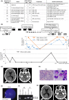
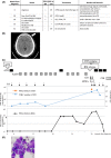
Central nervous system (CNS) involvement of acute promyelocytic leukemia (APL) causes poor prognosis. Our three cases show that CNS can be involved at the first hematological recurrence, but predicting this is difficult. Triple intrathecal treatment and craniospinal irradiation were effective, while arsenic oxide failed to prevent and improve CNS involvement.
Keywords: Acute promyelocytic leukemia; arsenic trioxide; central nervous system involvement; triple intrathecal treatment.
Figures


Similar articles
-
Acute Promyelocytic Leukemia Presenting with Cranial Nerve Involvement and Clivus Mass.Acta Haematol. 2025 Mar 29:1-7. doi: 10.1159/000545444. Online ahead of print. Acta Haematol. 2025. PMID: 40159328
-
Beneficial and adverse effects of molecularly targeted therapies for acute promyelocytic leukemia in central nervous system.CNS Neurol Disord Drug Targets. 2009 Nov;8(5):387-92. doi: 10.2174/187152709789541943. CNS Neurol Disord Drug Targets. 2009. PMID: 19702568 Review.
-
CNS relapses of acute promyelocytic leukemia after all-trans retinoic acid.Ann Pharmacother. 2002 Dec;36(12):1900-6. doi: 10.1345/aph.1A471. Ann Pharmacother. 2002. PMID: 12452754 Review.
-
The role of craniospinal irradiation in adults with a central nervous system recurrence of leukemia.Cancer. 2004 May 15;100(10):2176-80. doi: 10.1002/cncr.20280. Cancer. 2004. PMID: 15139061
-
Long-term outcome of relapsed acute promyelocytic leukemia treated with oral arsenic trioxide-based reinduction and maintenance regimens: A 15-year prospective study.Cancer. 2018 Jun 1;124(11):2316-2326. doi: 10.1002/cncr.31327. Epub 2018 Mar 26. Cancer. 2018. PMID: 29579321
Cited by
-
Acute Promyelocytic Leukemia: Update on the Mechanisms of Leukemogenesis, Resistance and on Innovative Treatment Strategies.Cancers (Basel). 2019 Oct 18;11(10):1591. doi: 10.3390/cancers11101591. Cancers (Basel). 2019. PMID: 31635329 Free PMC article. Review.
-
Intracranial acute promyelocytic leukemia at presentation-A case-based discussion.Leuk Res Rep. 2019 May 2;11:41-45. doi: 10.1016/j.lrr.2019.04.007. eCollection 2019. Leuk Res Rep. 2019. PMID: 31193772 Free PMC article.
References
-
- Sanz, M. A. , Vellenga E., Rayon C., Diaz‐Mediavilla J., Rivas C., Amutio E., et al. 2004. All‐trans retinoic acid and anthracycline monochemotherapy for the treatment of elderly patients with acute promyelocytic leukemia. Blood 104:3490–3493. - PubMed
-
- Sanz, M. A. , Martin G., Gonzalez M., Leon A., Rayon C., Rivas C., et al. 2004. Risk‐adapted treatment of acute promyelocytic leukemia with all‐trans‐retinoic acid and anthracycline monochemotherapy: a multicenter study by the PETHEMA group. Blood 103:1237–1243. - PubMed
-
- Lo‐Coco, F. , Avvisati G., Vignetti M., Breccia M., Gallo E., Rambaldi A., et al. 2010. Front‐line treatment of acute promyelocytic leukemia with AIDA induction followed by risk‐adapted consolidation for adults younger than 61 years: results of the AIDA‐2000 trial of the GIMEMA Group. Blood 116:3171–3179. - PubMed
-
- de Botton, S. , Sanz M. A., Chevret S., Dombret H., Martin G., Thomas X., et al. 2006. Extramedullary relapse in acute promyelocytic leukemia treated with all‐trans retinoic acid and chemotherapy. Leukemia 20:35–41. - PubMed
-
- Sanz, M. A. , Lo Coco F., Martin G., Avvisati G., Rayon C., Barbui T., et al. 2000. Definition of relapse risk and role of nonanthracycline drugs for consolidation in patients with acute promyelocytic leukemia: a joint study of the PETHEMA and GIMEMA cooperative groups. Blood 96:1247–1253. - PubMed
Publication types
LinkOut - more resources
Full Text Sources
Other Literature Sources

