Effective localization in tumor-induced osteomalacia using 68Ga-DOTATOC-PET/CT, venous sampling and 3T-MRI
- PMID: 28469928
- PMCID: PMC5409940
- DOI: 10.1530/EDM-17-0005
Effective localization in tumor-induced osteomalacia using 68Ga-DOTATOC-PET/CT, venous sampling and 3T-MRI
Abstract
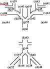
Summary: Tumor-induced osteomalacia (TIO) is a rare paraneoplastic syndrome characterized by renal phosphate wasting leading to hypophosphatemia due to excessive actions of fibroblast growth factor 23 (FGF23) produced by the tumors. Although the best way of curing TIO is complete resection, it is usually difficult to detect the culprit tumors by general radiological modalities owing to the size and location of the tumors. We report a case of TIO in which the identification of the tumor by conventional imaging studies was difficult. Nonetheless, a diagnosis was made possible by effective use of multiple modalities. We initially suspected that the tumor existed in the right dorsal aspect of the scapula by 68Ga-DOTATOC positron emission tomography/computed tomography (68Ga-DOTATOC-PET/CT) and supported the result by systemic venous sampling (SVS). The tumor could also be visualized by 3T-magnetic resonance imaging (MRI), although it was not detected by 1.5T-MRI, and eventually be resected completely. In cases of TIO, a stepwise approach of 68Ga-DOTATOC-PET/CT, SVS and 3T-MRI can be effective for confirmation of diagnosis.
Learning points: TIO shows impaired bone metabolism due to excessive actions of FGF23 produced by the tumor. The causative tumors are seldom detected by physical examinations and conventional radiological modalities.In TIO cases, in which the localization of the culprit tumors is difficult, 68Ga-DOTATOC-PET/CT should be performed as a screening of localization and thereafter SVS should be conducted to support the result of the somatostatin receptor (SSTR) imaging leading to increased diagnosability.When the culprit tumors cannot be visualized by conventional imaging studies, using high-field MRI at 3T and comparing it to the opposite side are useful after the tumor site was determined.
Figures


References
-
- Folpe AL, Fanburg-Smith JC, Billings SD, Bisceglia M, Bertoni F, Cho JY, Econs MJ, Inwards CY, Jan de Beur SM, Mentzel T, et al. 2004. Most osteomalacia-associated mesenchymal tumors are a single histopathologic entity: an analysis of 32 cases and a comprehensive review of the literature. American Journal of Surgical Pathology 28 1–30. ( 10.1097/00000478-200401000-00001) - DOI - PubMed
-
- Tarasova VD, Trepp-Carrasco AG, Thompson R, Recker RR, Chong WH, Collins MT, Armas LAG. 2013. Successful treatment of tumor-induced osteomalacia due to an intracranial tumor by fractionated stereotactic radiotherapy. Journal of Clinical Endocrinology and Metabolism 98 4267–4272. ( 10.1210/jc.2013-2528) - DOI - PMC - PubMed
LinkOut - more resources
Full Text Sources
Other Literature Sources

