. 2017 Apr 1;7(4):913-922.
eCollection 2017.

miR-381-3p suppresses the proliferation of oral squamous cell carcinoma cells by directly targeting FGFR2

Affiliations

miR-381-3p suppresses the proliferation of oral squamous cell carcinoma cells by directly targeting FGFR2

Xiao Yang et al. Am J Cancer Res. .

Abstract

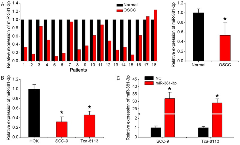
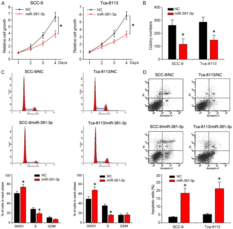
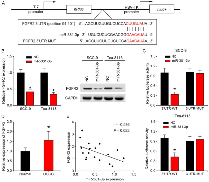
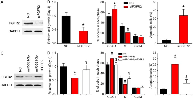
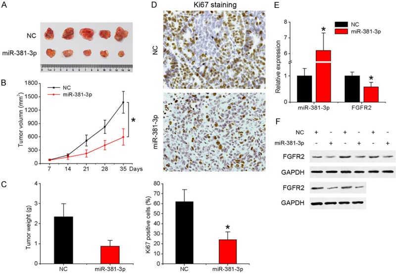
Mutiple microRNAs are implicated in oral squamous cell carcinoma (OSCC), which is characterized by a high rate of proliferation and nodal metastasis. Data from the present study showed that miR-381-3p is significantly underexpressed in both OSCC tissues and cell lines. Overexpression of miR-381-3p led to marked suppression of proliferation and cell cycle progression of OSCC cells and promotion of apoptosis. Notably, fibroblast growth factor receptor 2 (FGFR2) was downregulated by miR-381-3p through direct interactions with its 3' untranslated region. Knockdown of FGFR2 recapitulated the growth suppressive effect of miR-381-3p. Conversely, restoring FGFR2 expression attenuated miR-381-3p-induced effects in OSCC cells. Expression patterns of miR-381-3p and FGFR2 were inversely correlated in OSCC tissues. Our collective results provide novel evidence that miR-381-3p acts as a tumor suppressor in OSCC by directly targeting FGFR2, thereby presenting a promising therapeutic target.

Keywords: FGFR2; miR-381-3p; oral squamous cell carcinoma; proliferation.


Conflict of interest statement

None.

Figures

Figure 1
Figure 1
miR-381-3p expression in OSCC tissues and cell lines. A. Relative miR-381-3p expression in 18 pairs of OSCC specimens and matched adjacent normal specimens detected with qRT-PCR. U6 was used as an internal control. B. qRT-PCR analysis of miR-381-3p expression in OSCC cell lines (SCC-9 and Tca-8113) and HOK cells. C. SCC-9 and Tca-8113 cells were infected with miR-381-3p or negative control lentivirus, and qRT-PCR used to determine miR-381-3p expression. Data represent means ± SD of three independent experiments. *P < 0.05.
Figure 2
Figure 2
Effects of miR-381-3p on proliferation, cell cycle and apoptosis of OSCC cells. SCC-9 and Tca-8113 cells were infected with miR-381-3p or control lentivirus. A. Cell viability was detected with the MTT assay. B. Counting of colony numbers. C. Flow cytometry analysis of cell cycle progression. D. Apoptosis was measured via flow cytometry. Data represent means ± SD of three independent experiments. *P < 0.05.
Figure 3
Figure 3
FGFR2 is a direct downstream target of miR-381-3p. A. A fragment of FGFR2 3’-UTR containing the wild-type (WT) miR-381-3p or mutant binding site was cloned downstream of the reporter gene vector. B. Levels of FGFR2 mRNA and protein were measured using qRT-PCR and western blot, respectively, in response to overexpression of miR-381-3p in SCC-9 and Tca-8113 cells. C. Luciferase activity of wild-type or mutant FGFR2 3’-UTR luciferase vectors in the presence of miR-381-3p. D. FGFR2 mRNA expression in 18 pairs of tongue OSCC specimens and matched adjacent normal specimens. E. Scatter plot showing the correlation between miR-381-3p and FGFR2 expression in OSCC tissues. Data represent means ± SD of three independent experiments. *P < 0.05.
Figure 4
Figure 4
miR-381-3p suppresses OSCC growth by downregulating FGFR2. A. SCC-9 cells were transfected with FGFR2 siRNA or negative control. After 48 h, the FGFR2 protein level was examined via western blot analysis. B. MTT, cell cycle and apoptosis assays. C. SCC-9 cells were infected with NC, miR-381-3p lentivirus or miR-381-3p and FGFR2 lentivirus. After 48 h, FGFR2 protein levels were examined via western blot analysis. D. MTT, cell cycle and cell apoptosis assays. Data represent means ± SD of three independent experiments. *P < 0.05 vs. NC group, $P < 0.05 vs. miR-381-3p group.
Figure 5
Figure 5
Overexpression of miR-381-3p inhibits OSCC tumorigenesis in vivo. Tumors were detected every 7 days after implantation of SCC-9 cells infected with miR-381-3p or control lentivirus. A. Representative images of the tumors formed. B. Tumor volume. C. Mean weight. D. Ki-67-stained sections of transplanted tumors (×400). E. qRT-PCR analysis of expression of miR-381-3p and FGFR2 in tumor tissues. F. Western blot analysis of FGFR2 expression in tumors. Data represent means ± SD of three independent experiments. *P < 0.05.

References

    1. Warnakulasuriya S. Global epidemiology of oral and oropharyngeal cancer. Oral Oncol. 2009;45:309–16. - PubMed
    1. Sano D, Myers JN. Metastasis of squamous cell carcinoma of the oral tongue. Cancer Metastasis Rev. 2007;26:645–62. - PubMed
    1. Huntzinger E, Izaurralde E. Gene silencing by microRNAs: contributions of translational repression and mRNA decay. Nat Rev Genet. 2011;12:99–110. - PubMed
    1. Esquela-Kerscher A, Slack FJ. Oncomirs-microRNAs with a role in cancer. Nat Rev Cancer. 2006;6:259–69. - PubMed
    1. Kong D, Zhang G, Ma H, Jiang G. miR-1271 inhibits OSCC cell growth and metastasis by targeting ALK. Neoplasma. 2015;62:559–66. - PubMed

LinkOut - more resources