. 2017 Apr 19:7:130.
doi: 10.3389/fcimb.2017.00130. eCollection 2017.

Signaling Mediated by Toll- Like Receptor 5 Sensing of Pseudomonas aeruginosa Flagellin Influences IL-1β and IL-18 Production by Primary Fibroblasts Derived from the Human Cornea

Affiliations

Signaling Mediated by Toll- Like Receptor 5 Sensing of Pseudomonas aeruginosa Flagellin Influences IL-1β and IL-18 Production by Primary Fibroblasts Derived from the Human Cornea

Maria Del Mar Cendra et al. Front Cell Infect Microbiol. .

Abstract

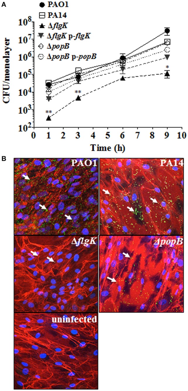
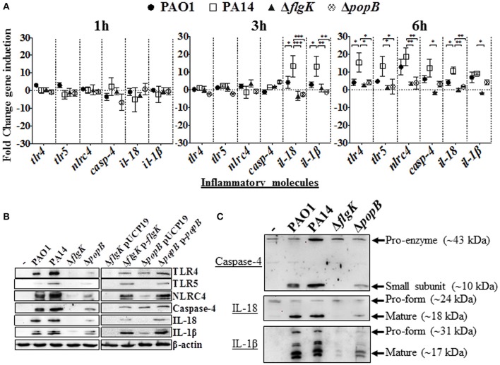
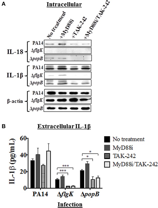
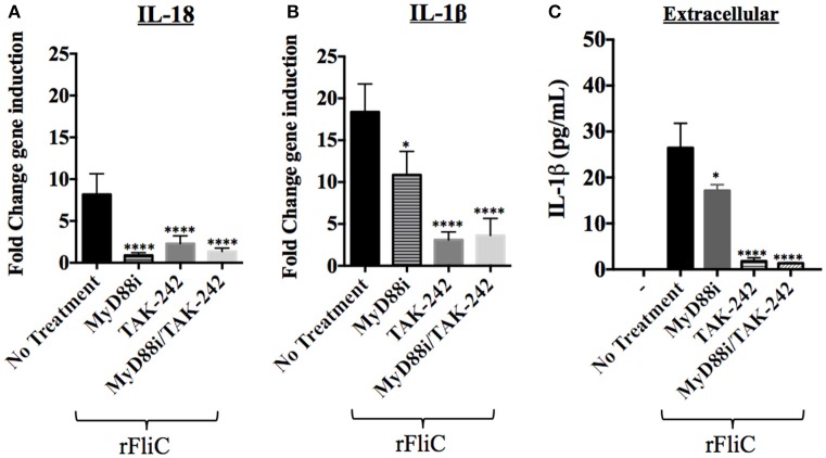
Pseudomonas aeruginosa is the principal cause of bacterial keratitis worldwide and overstimulation of the innate immune system by this organism is believed to contribute significantly to sight loss. In the current study, we have used primary human corneal fibroblast (hCF) cells as an ex vivo model of corneal infection to examine the role of P. aeruginosa flagellum and type three secretion system (TTSS) in inducing inflammasome-associated molecules that trigger IL-1β and IL-18 production during the early stages of the infection. Our results show that P. aeruginosa infection stimulated the non-canonical pathway for IL-1β and IL-18 expression and pathway stimulation was influenced predominantly by the flagellum. Both IL-1β and IL-18 cytokines were expressed intracellularly during bacterial infection, but only the former was released and detected in the extracellular environment. We also investigated the signaling pathways in hCFs mediated by Toll-Like Receptor (TLR)4 and TLR5 sensing of P. aeruginosa, and our data show that the signal triggered by TLR5-flagellin sensing significantly contributed to IL-1β and IL-18 cytokine production in our model. Our study suggests that IL-18 expression is wholly dependent on extracellular flagellin sensing by TLR5, whereas IL-1β expression is also influenced by P. aeruginosa lipopolysacharide. Additionally, we demonstrate that IL-1β and IL-18 production by hCFs can be triggered by both MyD88-dependent and -independent pathways. Overall, our study provides a rationale for the development of targeted therapies, by proposing an inhibition of flagellin-PRR-signaling interactions, in order to ameliorate the inflammatory response characteristic of P. aeruginosa keratitis.

Keywords: MyD88; NLRC4; Pseudomonas aeruginosa; TLR5; cytokine; fibroblasts; flagellin; human cornea.


Figures

Figure 1
Figure 1
Expression of flagellum contributes to P. aeruginosa adhesion to hCFs. (A) Cells were infected with MOI = 10 of PAO1, PA14, ΔflgK, ΔpopB and complemented PA14 ΔflgK p-flgK, PA14 ΔpopB p-popB strains and association to hCFs monolayers was quantified by viable counting after 1, 3, 6 and 9 h. The symbols represent the means and bars the standard error of the mean (SEM) of n = 3 independent experiments. PA14 ΔflgK and PA14 ΔpopB strains carrying the cloning vector pUCP19 without insert were tested in order to exclude possible vector interferences: no differences were observed when compared with their respective mutant strains in their association capabilities (data not shown). Statistical significance was calculated with a one-way ANOVA with Dunnett's multiple comparison test and it is represented by asterisks as follows: *P < 0.05 and **P < 0.01. (B) Confocal microscopy images of GFP-expressing PAO1, PA14 and mutant strains after 9 h of infection. Arrows denote the positions of bacteria and images are representative of n = 3 independent experiments.
Figure 2
Figure 2
Inflammatory responses of human corneal fibroblast cells (hCFs) toward P. aeruginosa infection. (A) RT-qPCR of immune signaling molecules after 1, 3 and 6 h hCFs infection with PAO1 and PA14 wild-type and PA14 ΔflgK and PA14 ΔpopB mutant strains at a MOI = 10. Symbols represent the mean and bars the SEM of the log of gene expression from infected hCFs compared with uninfected controls from n = 10 independent hCF samples. Validation of the oligonucleotides used for RT-qPCR examination of gene expression in infected hCFs was done by demonstrating the same signals in THP-1 cells stimulated with 100 ng/mL of P. aeruginosa LPS (data not shown). Statistical significance was calculated with a one-way ANOVA with Dunnett's multiple comparison test and it is represented by asterisks as follows: *P < 0.05, **P < 0.01, ***P < 0.001. (B) Cells infected with MOI = 10 of PAO1 and PA14 wild-type, PA14 ΔflgK and PA14 ΔpopB mutant strains, and the complemented PA14 ΔflgK p-flgK, PA14 ΔpopB p-popB and control PA14 ΔflgK pUCP19 and PA14 ΔpopB pUCP19 strains were lysed and intracellular protein levels of TLR4, TLR5, NLRC4, active caspase-4 and matured forms of IL-18 and IL-1β were detected by western blot. Western blot image is representative of three independent experiments. (C) Western blot images show protein levels of pro- and matured forms of caspase-4, IL-18 and IL-1β in hCFs infected with PAO1 and PA14 wild-type and PA14 ΔflgK and PA14 ΔpopB mutant strains. Protein expression for uninfected hCFs is also shown. The rabbit polyclonal anti- caspase-4, -IL-18, and -IL-1β antibodies are specific for binding to the mature form of each respective protein and with less affinity for the pro-forms. Respective molecular weights of pro and mature forms are denoted with arrows. A representative image from n = 5 independent experiments is shown.
Figure 3
Figure 3
Extracellular levels of IL-18 and IL-1β cytokines during hCFs infection with P. aeruginosa. IL-1β and IL-18 cytokine release was measured after 6 h hCFs infection with PAO1 and PA14 wild-type and PA14 ΔflgK and PA14 ΔpopB mutant and complemented strains (MOI = 10). The columns represent the mean and error bars the SEM from n = 3 independent experiments. MSD-ELISA was used to quantify the levels of extracellular mature IL-1β (A) and IL-18 (B). Statistical significance was calculated with a one-way ANOVA with Dunnett's multiple comparison test and it is represented by asterisks as follows: ***P < 0.001. (C) Extracellular IL-18 detected by western blot in TCA-precipitated supernatants after 6 h infection of hCFs with P. aeruginosa wild-type and mutant strains. A total protein extract from hCFs infected with PA14 was used as a positive control for IL-18 detection (Control).
Figure 4
Figure 4
Effects of pathway inhibition on IL-18 and IL-1β production. Cells were treated with MyD88i, TAK-242, MyD88i/TAK-242 inhibitors and then infected with PAO1, PA14, ΔflgK and ΔpopB strains (MOI = 10). (A) Western blot was used to detect intracellular mature IL-18 and IL-1β cytokines after 6 h of bacterial infection. Detection of intracellular levels of β-actin was used as endogenous and loading control. (B) Quantification of extracellular IL-1β cytokine after 6 h of infection. The columns represent the means and the error bars the SEM of n = 3 independent experiments. Statistical significance was calculated with a one-way ANOVA with Dunnett's multiple comparison test and it is represented by asterisks as follows: *P < 0.05, ***P < 0.001.
Figure 5
Figure 5
Effect of TLR5 silencing in hCFs on IL-18 and IL-1β expression and release. Wild-type hCFs and siTLR5-hCFs were infected with PA14 wild-type and ΔflgK mutant strain at MOI = 10, and stimulated with 5 μg/mL of rFliC for 6 h. LAL assay quantified that rFliC contained ~150 pg of LPS/mg of protein (<0.00002% w/w LPS) and RT-qPCR showed that these levels of E. coli LPS did not stimulate any gene transcription in hCFs (data not shown). Non-infected monolayers were used as a control. (A,B) Gene expression of IL-18 (A) and IL-1β (B) was measured by RT-qPCR. The bars show the fold changes in gene expression in siTLR5-hCFs compared to wild-type hCFs, which were determined using the 2−ΔΔCT method (Livak and Schmittgen, 2001). (C) Extracellular IL-1β levels measured by MSD-ELISA. rFliC was delivered intracellularly using SLO treatment. SLO treatment alone did not stimulate significant IL-1β release by hCFs (data not shown). The graph shows the levels of extracellular IL-1β quantified in the supernatants of hCFs and siTLR5-hCFs after 6 h following the different infection and stimulatory conditions. Statistical significance was calculated with a one-way ANOVA with Dunnett's multiple comparison test and it is represented by asterisks as follows: *P < 0.05, ****P < 0.0001.
Figure 6
Figure 6
Effect of PA14 ΔflgK mutant LPS on IL-1β expression by hCFs. Wild-type hCFs and siTLR5-hCFs monolayers were treated with the TAK-242 inhibitor and then infected with PA14 ΔflgK mutant strain at MOI = 10 for 6 h. Un-treated and uninfected monolayers were used as control. (A) Gene expression of IL-1β was measured by RT-qPCR. The graph shows the fold changes of IL-1β expression calculated relative to the corresponding un-infected cell monolayer (hCF or siTLR5-hCF). (B) Intracellular IL-1β mature protein measured by western blot. A total protein extract from hCFs infected with PA14 was used as a positive control for IL-1β detection and intracellular levels of β-actin detected as an endogenous and loading control. (C) Gene expression of IL-1β in hCF monolayers un-treated and treated with TAK-242, Myd88i and TAK-242/Myd88i followed by stimulation with 100 ng/mL of P. aeruginosa LPS. The fold changes of IL-1β expression are calculated relative to un-stimulated monolayers. Statistical significance was calculated with a one-way ANOVA with Dunnett's multiple comparison test and it is represented by asterisks as follows: *P < 0.05, ****P < 0.0001.
Figure 7
Figure 7
Effect of MyD88i, TAK-242 and MyD88i/TAK-242 inhibition on IL-18 and IL-1β expression in hCF stimulated with rFliC. IL-18 and IL-1β gene expression was measured by RT-qPCR from rFliC-stimulated hCF following treatment with MyD88i, TAK-242 and MyD88i/TAK-242 and untreated cells. The graph shows the fold changes of IL-18 (A) and IL-1β (B) gene expression calculated relative to the unstimulated and untreated hCF monolayers. The significance of the changes in gene expression shown in each inhibitory condition is compared to that measured in uninhibited hCF stimulated with P. aeruginosa rFliC. (C) Extracellular levels of IL-1β quantified from supernatants of inhibited hCFs following stimulation with rFliC. SLO-treatment was used to deliver rFliC intracellularly and no significant extracellular IL-1β was detected by treatment with SLO alone (data not shown). Unstimulated and inhibited cells were used as controls (-). Statistical significance was calculated with a one-way ANOVA with Dunnett's multiple comparison test and it is represented by asterisks as follows: *P < 0.05, ****P < 0.0001.

References

    1. Auerbuch V., Golenbock D. T., Isberg R. R. (2009). Innate immune recognition of Yersinia pseudotuberculosis type III secretion. PLoS Pathog. 5:e1000686. 10.1371/journal.ppat.1000686 - DOI - PMC - PubMed
    1. Berg H. C. (2003). The rotary motor of bacterial flagella. Annu. Rev. Biochem. 72, 19–54. 10.1146/annurev.biochem.72.121801.161737 - DOI - PubMed
    1. Casson C. N., Yu J., Reyes V. M., Taschuk F. O., Yadav A., Copenhaver A. M., et al. (2015). Human caspase-4 mediates non-canonical inflammasome activation against gram-negative bacterial pathogens. Proc. Natl. Acad. Sci. U.S.A. 112, 6688–6693. 10.1073/pnas.1421699112 - DOI - PMC - PubMed
    1. Chen G., Shaw M. H., Kim Y. G., Nuñez G. (2009). NOD-like receptors: role in innate immunity and inflammatory disease. Annu. Rev. Pathol. 4, 365–398. 10.1146/annurev.pathol.4.110807.092239 - DOI - PubMed
    1. Choi Y. J., Im E., Chung H. K., Pothoulakis C., Rhee S. H. (2010). TRIF mediates Toll-like receptor 5-induced signaling in intestinal epithelial cells. J. Biol. Chem. 285, 37570–37578. 10.1074/jbc.M110.158394 - DOI - PMC - PubMed

MeSH terms

LinkOut - more resources