. 2017 Apr 26;3(4):329-337.
doi: 10.1021/acscentsci.7b00026. Epub 2017 Feb 24.

In Vivo Activation of Duocarmycin-Antibody Conjugates by Near-Infrared Light

Affiliations

In Vivo Activation of Duocarmycin-Antibody Conjugates by Near-Infrared Light

Roger R Nani et al. ACS Cent Sci. .

Abstract

Near-IR photocaging groups based on the heptamethine cyanine scaffold present the opportunity to visualize and then treat diseased tissue with potent bioactive molecules. Here we describe fundamental chemical studies that enable biological validation of this approach. Guided by rational design, including computational analysis, we characterize the impact of structural alterations on the cyanine uncaging reaction. A modest change to the ethylenediamine linker (N,N'-dimethyl to N,N'-diethyl) leads to a bathochromic shift in the absorbance maxima, while decreasing background hydrolysis. Building on these structure-function relationship studies, we prepare antibody conjugates that uncage a derivative of duocarmycin, a potent cytotoxic natural product. The optimal conjugate, CyEt-Pan-Duo, undergoes small molecule release with 780 nm light, exhibits activity in the picomolar range, and demonstrates excellent light-to-dark selectivity. Mouse xenograft studies illustrate that the construct can be imaged in vivo prior to uncaging with an external laser source. Significant reduction in tumor burden is observed following a single dose of conjugate and near-IR light. These studies define key chemical principles that enable the identification of cyanine-based photocages with enhanced properties for in vivo drug delivery.


Conflict of interest statement

The authors declare no competing financial interest.

Figures
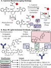
Figure 1
Figure 1
(A) Mechanism of the cyanine uncaging reaction and (B) evolution of the near-IR light-activated ADC strategy.
Figure 2
Figure 2
Impact of modifications to the cyanine caging scaffold on photooxidation efficiency (method A), uncaging kinetics (method B), and background hydrolysis (method C). Method A: Absorbance traces at the λmax of a 1 μM solution of each cyanine cage in pH 7.4 PBS irradiated with a 690 ± 20 nm LED (20 mW/cm2). Method B: Umb release (measured from a standard curve using fluorescence) from cyanine cage (1 μM, pH 7.4 PBS). Exhaustive photooxidation at 0 °C (15–180 J/cm2) was followed by monitoring Umb release at 37 °C (30 min intervals). Method C: Umb release at 37 °C (2.5 h intervals) from cyanine cage (10 μM, pH 7.4 PBS). For methods A and B, the experiments were run to completion and the data was fit to one phase decay parameters. For method C, the slow rate of background Umb release precluded this analysis. The reported values for the time to 10% reaction conversion (t10%) is either directly observed or extrapolated from the initial slope (see Supporting Information for details). *Less than 20% Umb release yield after 5 h.
Scheme 1
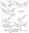
Scheme 1. Synthesis of Cyanines 18 and 19
Figure 3
Figure 3
Effect of light dose and wavelength on the in vitro efficacy of CyMe-Pan-Duo and CyEt-Pan-Duo. (A) The relationship between absorbance spectra and the irradiation wavelength. (B) MDA-MB-468 (EGFR+) cell viability under control conditions (vehicle, free duocarmycin DM (Duo DM), and unirradiated ADC). (C, D) Cell viability as a function of irradiation light dose and wavelength in the presence of 100 pM of each ADC (solid line, CyMe-Pan-Duo; dashed line, CyEt-Pan-Duo). Error bars represent standard deviation (n = 4).
Figure 4
Figure 4
In vivo efficacy of CyEt-Pan-Duo (DOL 4) in MDA-MB-468-luc tumor-bearing mice. (A) Conjugate dosing (i.v.), irradiation (690 nm, 80 J/cm2, 800 mW/cm2), and imaging regimen. (B) Fluorescence images at 800 nm. (C) Bioluminescence images of luciferase activity. (D) Luciferase activity as a function of time post-irradiation, relative to initial. (E) Tumor volume as a function of time post-irradiation. (F) Survival as a function of time post-irradiation. For D–F, vehicle (black), 100 μg of CyEt-Pan-Duo – hν (red), and 10 μg (green), 30 μg (purple), and 100 μg (orange) of CyEt-Pan-Duo + hν. n = 9 mice per condition. *p < 0.05, **p < 0.01, Dunnett’s test with ANOVA (D, E) with error bars representing standard error of the mean (SEM) or log-rank test (F).

References

    1. Nguyen Q. T.; Tsien R. Y. Fluorescence-Guided Surgery with Live Molecular Navigation--A New Cutting Edge. Nat. Rev. Cancer 2013, 13, 653–662. 10.1038/nrc3566. - DOI - PMC - PubMed
    1. Garland M.; Yim J. J.; Bogyo M. A Bright Future for Precision Medicine: Advances in Fluorescent Chemical Probe Design and Their Clinical Application. Cell Chem. Biol. 2016, 23, 122–136. 10.1016/j.chembiol.2015.12.003. - DOI - PMC - PubMed
    1. Dolmans D. E.; Fukumura D.; Jain R. K. Photodynamic Therapy for Cancer. Nat. Rev. Cancer 2003, 3, 380–387. 10.1038/nrc1071. - DOI - PubMed
    1. Mallidi S.; Anbil S.; Bulin A. L.; Obaid G.; Ichikawa M.; Hasan T. Beyond the Barriers of Light Penetration: Strategies, Perspectives and Possibilities for Photodynamic Therapy. Theranostics 2016, 6, 2458–2487. 10.7150/thno.16183. - DOI - PMC - PubMed
    1. Lerch M. M.; Hansen M. J.; van Dam G. M.; Szymanski W.; Feringa B. L. Emerging Targets in Photopharmacology. Angew. Chem., Int. Ed. 2016, 55, 10978–10999. 10.1002/anie.201601931. - DOI - PubMed