Comparative Study
. 2017 Aug;40(8):1237-1245.
doi: 10.1007/s00270-017-1659-4. Epub 2017 May 3.

Continuously Grooved Stent Struts for Enhanced Endothelial Cell Seeding

Affiliations
Comparative Study

Continuously Grooved Stent Struts for Enhanced Endothelial Cell Seeding

Marja Ter Meer et al. Cardiovasc Intervent Radiol. 2017 Aug.

Abstract

Purpose: Implantation of pre-endothelialized stents could enhance cellular recovery of a damaged vessel wall provided attached cells remain viable, functional and are present in sufficient numbers after deployment. The purpose of this study was to evaluate the feasibility of grooved stainless steel (SS) stents as a primary endothelial cell (EC) carrier with potentially enhanced EC protection upon stent deployment.

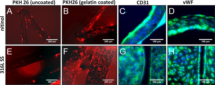
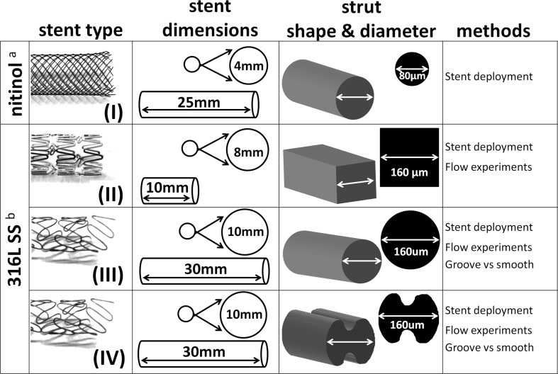
Materials and methods: Attachment and behavior of enzymatically harvested human adult venous ECs seeded onto gelatin-coated vascular stents were evaluated in an in vitro setting. Smooth and grooved SS stents and smooth nitinol stents were studied.

Results: All cells expressed EC markers vWF and CD31. Using rotational seeding for a period of 16-24 h, ECs attached firmly to the stents with sufficient coverage to form a confluent EC monolayer. The grooved SS wire design was found to enable attachment of three times the number of cells compared to smooth wires. This also resulted in an increased number of cells remaining on the stent after deployment and after pulsatile flow of 180 ml/min for 24 h, which did not result in additional EC detachment.

Conclusions: The grooved stent provides a potential percutaneous means to deliver sufficient numbers of viable and functional cells to a vessel segment during vascular intervention. The grooves were found to offer a favorable surface for EC attachment and protection during stent deployment in an in vitro setting.

Keywords: 316L stainless steel; Cell adhesion; Endothelialization; Endovascular stent; In vitro; Nitinol.


Conflict of interest statement

Conflict of interest

On behalf of all authors, the corresponding author states that there is no conflict of interest.

Human and Animal Rights

All procedures performed in studies involving human participants were in accordance with the ethical standards of the institutional and/or national research committee and with the 1964 Helsinki declaration and its later amendments or comparable ethical standards.

Figures

Fig. 1
Fig. 1
Summary of preliminary experiments: Optimization of surface coating using gelatin for EC seeding onto stents and characterization of attached ECs. Attachment of ECs to gelatin-coated stents, both nitinol and 316L SS, was superior over attachment to uncoated stents after a 24 h seeding period (A, B, E, F). Cells were stained with a fluorescent membrane marker PKH26 for visualization. Attached cells had a proper phenotype as EC-specific markers von Willebrand factor and CD31 (PECAM-1) were expressed (both green), cells were counterstained with DAPI in blue (C, D, G, F)
Fig. 2
Fig. 2
An overview of the stent design and dimensions of stents and struts used in the current study. aSelf-expanding, bballoon expandable
Fig. 3
Fig. 3
Stent deployment resulted in a loss of seeded EC for all stents, subsequent exposure to flow did not result in additional cell loss. A Stent (I) deployment by pulling the stent through a small tube. B Stent (II) deployment inside a PharMed tube using either an angioplasty balloon (top) or a pipette tip (bottom). C The tubular glass chamber used for flow experiments. D The stent outline is highlighted to demonstrate loss of cells on the lateral side of stent (II). E Due to stent deployment, a cell loss of over 50% of the cells was observed based on a CCK-8 assay. F The CCK-8 assay confirmed there was minimal additional cell loss from the stents (II) after exposure to flow (2.0 ± 2.2%; n = 5)
Fig. 4
Fig. 4
Stents (IV) consisting of 316L wire afforded with grooves can accommodate three times more cells compared to a 316L stent (III) with the same size and the same design but without grooves. A SEM images of the smooth and grooved stent struts. B SEM image showing the continuous helical groove. C Groove design, ∅ 40-µm grooves resulting in an area increase of 20%. D Cells were labeled with PKH26, cells covered the entire surface of the smooth stent. E A very dense cell population was observed within the grooves compared to the rest of the surface. F Based on the CCK-8 assay, the grooved stent could accommodate 3.2 ± 1.4 times as many cells as the smooth stent, and the relative cell count after deployment was also higher for the grooved stent
Fig. 5
Fig. 5
ECs seeded onto gelatin-coated stents (I) and (III) are still proliferative and able to endothelialize a surface beyond the stent surface. A, B phase contrast images after 48 h in culture

References

    1. Inoue T, Croce K, Morooka T, Sakuma M, Node K, Simon DI. Vascular inflammation and repair: implications for re-endothelialization, restenosis, and stent thrombosis. JACC Cardiovasc Interv. 2011;4(10):1057–1066. doi: 10.1016/j.jcin.2011.05.025. - DOI - PMC - PubMed
    1. Richter GM, Palmaz JC, Noeldge G, Tio F. Relationship between blood flow, thrombus, and neointima in stents. J Vasc Interv Radiol. 1999;10(5):598–604. doi: 10.1016/S1051-0443(99)70090-4. - DOI - PubMed
    1. Kipshidze N, Dangas G, Tsapenko M, Moses J, Leon MB, Kutryk M, et al. Role of the endothelium in modulating neointimal formation: vasculoprotective approaches to attenuate restenosis after percutaneous coronary interventions. J Am Coll Cardiol. 2004;44:733–739. - PubMed
    1. Douglas G, Van Kampen E, Hale AB, McNeill E, Patel J, Crabtree MJ, et al. Endothelial cell repopulation after stenting determines in-stent neointima formation: effects of bare-metal versus drug-eluting stents and genetic endothelial cell modification. Eur Heart J. 2012;34(43):3378–3388. doi: 10.1093/eurheartj/ehs240. - DOI - PMC - PubMed
    1. Tesfamariam B. Endothelial repair and regeneration following intimal injury. J Cardiovasc Transl Res. 2016;9(2):91–101. doi: 10.1007/s12265-016-9677-1. - DOI - PubMed

Publication types

LinkOut - more resources