

Bovine myoblast cell production in a microcarriers-based system
- PMID: 28470539
- PMCID: PMC5851947
- DOI: 10.1007/s10616-017-0101-8
Bovine myoblast cell production in a microcarriers-based system
Abstract
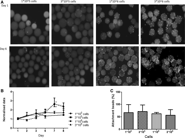
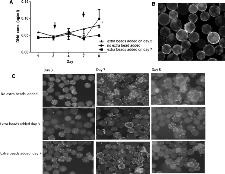
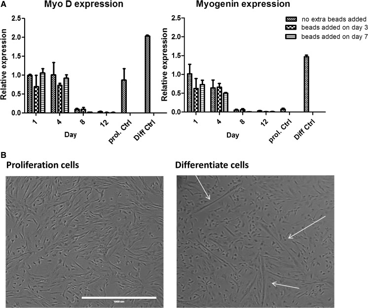
For several tissue engineering applications, in particular food products, scaling up culture of mammalian cells is a necessary task. The prevailing method for large scale cell culture is the stirred tank bioreactor where anchor dependent cells are grown on microcarriers suspended in medium. We use a spinner flask system with cells grown on microcarriers to optimize the growth of bovine myoblasts. Freshly isolated primary cells were seeded on microcarriers (Synthemax®, CellBIND® and Cytodex® 1 MCs). In this study, we provide proof of principle that bovine myoblasts can be cultured on microcarriers. No major differences were observed between the three tested microcarriers, except that sparsely populated beads were more common with CellBIND® and Synthemax® II beads suggesting a slower initiation of exponential growth than on Cytodex®. We also provide direct evidence that bovine myoblasts display bead-to-bead transfer. A remarkable pick up of growth was observed by adding new MCs. Bovine myoblasts seem to behave like human mesenchymal stem cells. Thus, our results provide valuable data to further develop and scale-up the production of bovine myoblasts as a prerequisite for efficient and cost-effective development of cultured meat. Applicability to other anchorage dependent cells can extend the importance of these results to cell culture for medical tissue engineering or cell therapy.
Keywords: Bioreactor; Cell culture; Microcarriers; Myoblast.
Figures

Similar articles
-
Attachment promoting compounds significantly enhance cell proliferation and purity of bovine satellite cells grown on microcarriers in the absence of serum.Front Bioeng Biotechnol. 2024 Nov 1;12:1443914. doi: 10.3389/fbioe.2024.1443914. eCollection 2024. Front Bioeng Biotechnol. 2024. PMID: 39553395 Free PMC article.
-
In vitro culture of primary human myoblasts by using the dextran microcarriers Cytodex3®.Folia Histochem Cytobiol. 2016;54(2):81-90. doi: 10.5603/FHC.a2016.0010. Epub 2016 Jun 8. Folia Histochem Cytobiol. 2016. PMID: 27270505
-
Biodegradable poly-ε-caprolactone microcarriers for efficient production of human mesenchymal stromal cells and secreted cytokines in batch and fed-batch bioreactors.Cytotherapy. 2017 Mar;19(3):419-432. doi: 10.1016/j.jcyt.2016.11.009. Epub 2016 Dec 22. Cytotherapy. 2017. PMID: 28017598
-
Microcarriers for Upscaling Cultured Meat Production.Front Nutr. 2020 Feb 20;7:10. doi: 10.3389/fnut.2020.00010. eCollection 2020. Front Nutr. 2020. PMID: 32154261 Free PMC article. Review.
-
Functional cells cultured on microcarriers for use in regenerative medicine research.Cell Transplant. 2011;20(1):49-62. doi: 10.3727/096368910X532792. Epub 2010 Sep 30. Cell Transplant. 2011. PMID: 20887678 Review.
Cited by
-
The important role of cellular mechanical microenvironment in engineering structured cultivated meat: Recent advances.Curr Res Food Sci. 2024 Sep 21;9:100865. doi: 10.1016/j.crfs.2024.100865. eCollection 2024. Curr Res Food Sci. 2024. PMID: 39416367 Free PMC article. Review.
-
Processing of fast-gelling hydrogel precursors in microfluidics by electrocoalescence of reactive species.Soft Matter. 2021 Nov 24;17(45):10312-10321. doi: 10.1039/d1sm01176f. Soft Matter. 2021. PMID: 34664052 Free PMC article.
-
Conceptual evolution and scientific approaches about synthetic meat.J Food Sci Technol. 2020 Jun;57(6):1991-1999. doi: 10.1007/s13197-019-04155-0. Epub 2019 Nov 14. J Food Sci Technol. 2020. PMID: 32431325 Free PMC article. Review.
-
Technical requirements for cultured meat production: a review.J Anim Sci Technol. 2021 Jul;63(4):681-692. doi: 10.5187/jast.2021.e45. Epub 2021 Jul 31. J Anim Sci Technol. 2021. PMID: 34447948 Free PMC article. Review.
-
Emerging technologies towards extracellular vesicles large-scale production.Bioact Mater. 2025 Jun 13;52:338-365. doi: 10.1016/j.bioactmat.2025.06.005. eCollection 2025 Oct. Bioact Mater. 2025. PMID: 40585384 Free PMC article. Review.
References
LinkOut - more resources
Full Text Sources
Other Literature Sources
