. 2017 Aug;26(8):1667-1673.
doi: 10.1002/pro.3184. Epub 2017 May 12.

Trapped intermediate state of plant pyruvate phosphate dikinase indicates substeps in catalytic swiveling domain mechanism

Affiliations

Trapped intermediate state of plant pyruvate phosphate dikinase indicates substeps in catalytic swiveling domain mechanism

Alexander Minges et al. Protein Sci. 2017 Aug.

Abstract

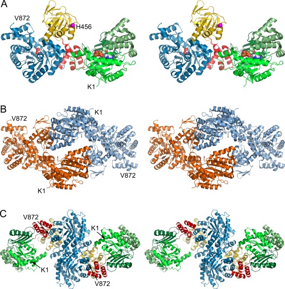
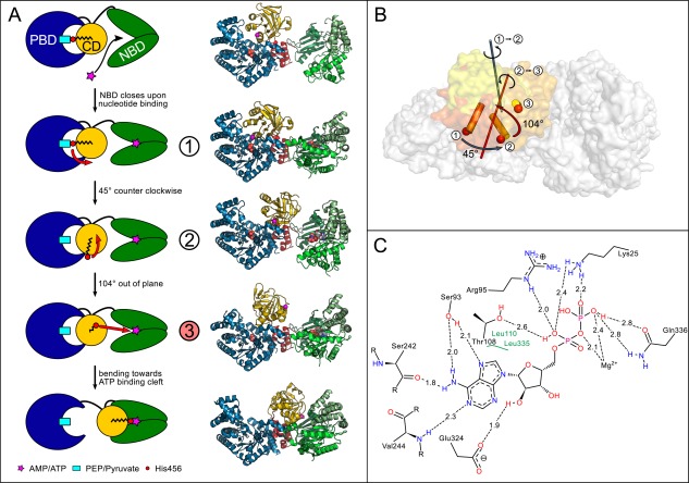
Pyruvate phosphate dikinase (PPDK) is an essential enzyme of both the C4 photosynthetic pathway and cellular energy metabolism of some bacteria and unicellular protists. In C4 plants, it catalyzes the ATP- and Pi -dependent formation of phosphoenolpyruvate (PEP) while in bacteria and protozoa the ATP-forming direction is used. PPDK is composed out of three distinct domains and exhibits one of the largest single domain movements known today during its catalytic cycle. However, little information about potential intermediate steps of this movement was available. A recent study resolved a discrete intermediate step of PPDK's swiveling movement, shedding light on the details of this intriguing mechanism. Here we present an additional structural intermediate that possibly represents another crucial step in the catalytic cycle of PPDK, providing means to get a more detailed understanding of PPDK's mode of function.

Keywords: C4 photosynthesis; catalytic intermediate; pyruvate phosphate dikinase; swiveling domain mechanism.


Figures

Figure 1
Figure 1
(A) Stereo cartoon representation of 5LU4 chain A illustrating the overall domain organization. The nucleotide binding domain (NBD, aa 1–340) and its three subdomains are colored in different greens. The PEP/pyruvate binding domain (PBD) is colored in blue (aa 534–872). The central domain (CD, yellow, aa 381–516) with the catalytic His456 (magenta, Cα shown as sphere) is attached to both substrate binding domains via two short linker helices (red, aa 341–380 and 517–533). Pyruvate and ADP bound to the PBD and NBD respectively are depicted as spheres. (B) Dimeric assembly within the asymmetric unit (ASU). The dimer is formed by contacts between the NBDs and CDs of chains A and B, colored orange and blue respectively. (C) Biological assembly as identified by the program EPPIC 11 and reconstructed from crystal symmetry. The dimerization interface is formed by both PBDs as previously described 12. Individual domains are colored according to (A). An interactive view is available in the electronic version of the article
Figure 2
Figure 2
(A) Schematic model of the CD movement in the catalytic cycle taking into account currently known conformational intermediates. Helix 20 containing the catalytic His456 (red circle) at its N‐terminal end is drawn as black zig‐zag structure. The different CD conformations are numbered according to (B) with the newly solved intermediate structure highlighted in red. Corresponding crystal structures are shown on the right from top to bottom: 5JVJ chain A, 5JVL chain D, 5JVN, 5LU4 chain A 6, 1KBL 7. (B) Illustration of PPDK‐CD structural intermediates. CDs of published structures 5JVL chain C (1, dark‐orange), 5JVN (2, orange), and of newly resolved structure 5LU4 (3, yellow) as well as the corresponding rotational axes for transformation between the different intermediate states are shown. (C) Schematic representation of the nucleotide binding site in 5LU4 chain A containing tightly bound ADP. The distances between the bound nucleotide and interacting amino acids of the binding pocket are given in Å. An interactive view is available in the electronic version of the article

References

    1. Mertens E (1993) ATP versus pyrophosphate: glycolysis revisited in parasitic protists. Parasitol Today 9:122–126. doi:10.1016/0169-4758(93)90169-g. - DOI - PubMed
    1. Chastain CJ, Chollet R (2003) Regulation of pyruvate, orthophosphate dikinase by ADP‐/pi‐dependent reversible phosphorylation in C3 and C4 plants. Plant Physiol Biochem 41:523–532. doi:10.1016/s0981-9428(03)00065-2. - DOI
    1. Nakanishi T, Nakatsu T, Matsuoka M, Sakata K, Kato H (2005) Crystal structures of pyruvate phosphate dikinase from maize revealed an alternative conformation in the swiveling‐domain motion. Biochemistry 44:1136–1144. doi:10.1021/bi0484522. - DOI - PubMed
    1. Cosenza LW, Bringaud F, Baltz T, Vellieux FM The 3.0 Å resolution crystal structure of glycosomal pyruvate phosphate dikinase from Trypanosoma brucei . J Mol Biol 318:1417–1432. doi:10.1016/s0022-2836(02)00113-4. - DOI - PubMed
    1. Minges A, Ciupka D, Winkler C, Höppner A, Gohlke H, Groth G (2017) Structural intermediates and directionality of the swiveling motion of pyruvate phosphate dikinase. Sci Rep 7:45389. doi:10.1038/srep45389. - DOI - PMC - PubMed

MeSH terms

LinkOut - more resources