

Loss of SH3GL2 promotes the migration and invasion behaviours of glioblastoma cells through activating the STAT3/MMP2 signalling
- PMID: 28470949
- PMCID: PMC5661104
- DOI: 10.1111/jcmm.13184
Loss of SH3GL2 promotes the migration and invasion behaviours of glioblastoma cells through activating the STAT3/MMP2 signalling
Abstract
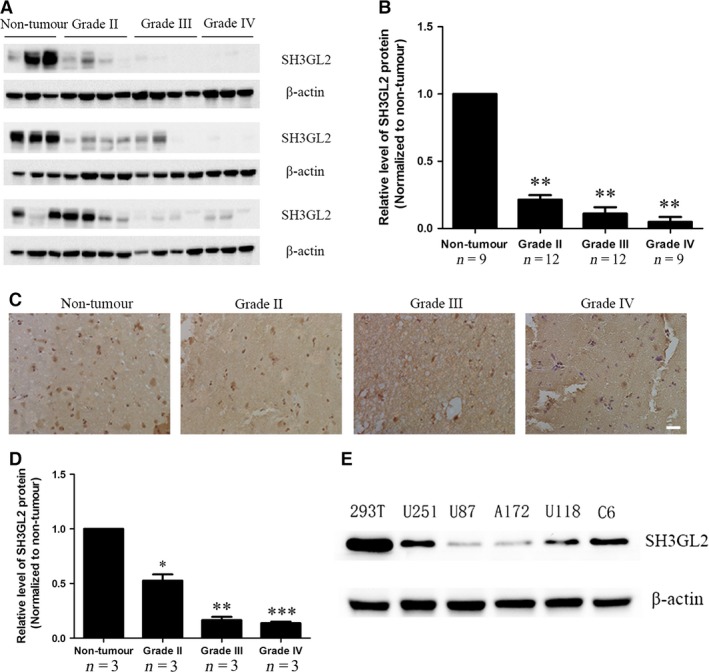
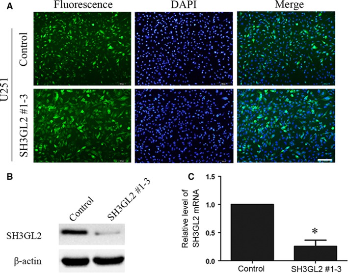
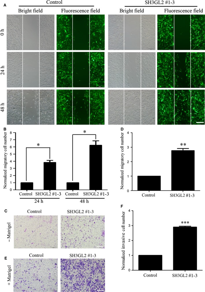
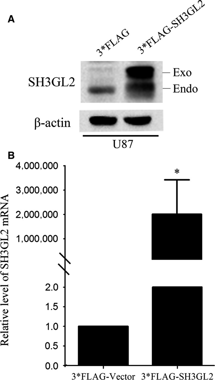
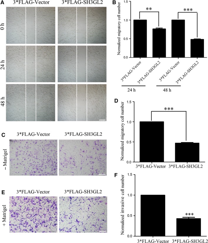
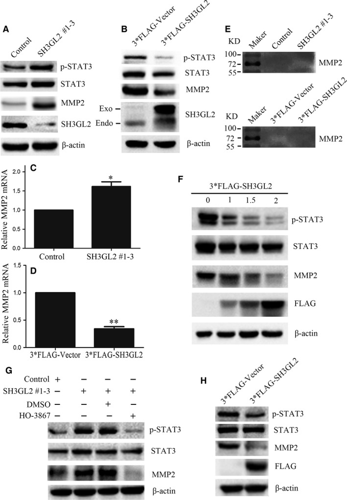
SH3GL2 (Src homology 3 (SH3) domain GRB2-like 2) is mainly expressed in the central nervous system and regarded as a tumour suppressor in human glioma. However, the molecular mechanism of the SH3GL2 protein involved in malignant behaviours of human glioma has not been elucidated. In this study, we tried to investigate the role of SH3GL2 in glioma cell migration and invasion and explore its underlined molecular mechanism. Firstly, we discovered that the protein level of SH3GL2 was widely decreased in the human glioma patients, especially in high-grade glioma tissues. Then, we determined the role of SH3GL2 in migration and invasion of glioma cells upon SH3GL2 knocking down and overexpressing. It was showed that knocking down of SH3GL2 promoted the migration and invasion of glioma cells, whereas overexpression of SH3GL2 inhibited them. Further study on molecular mechanism disclosed that silencing of SH3GL2 obviously activated the STAT3 (signal transducer and activator of transcription 3) signalling thereby promoting the expression and secretion of MMP2. On the contrary, overexpression of SH3GL2 had opposite effect. Taken together, the above results suggest that SH3GL2 suppresses migration and invasion behaviours of glioma cells through negatively regulating STAT3/MMP2 signalling and that loss of SH3GL2 may intensify the STAT3/MMP2 signalling thereby contributing to the migration and invasion of glioma cells.
Keywords: MMP2; SH3GL2; STAT3; glioma.
© 2017 The Authors. Journal of Cellular and Molecular Medicine published by John Wiley & Sons Ltd and Foundation for Cellular and Molecular Medicine.
Figures
Similar articles
-
IL-6 augments the invasiveness of U87MG human glioblastoma multiforme cells via up-regulation of MMP-2 and fascin-1.Oncol Rep. 2010 Jun;23(6):1553-9. doi: 10.3892/or_00000795. Oncol Rep. 2010. PMID: 20428809
-
MiR-330-mediated regulation of SH3GL2 expression enhances malignant behaviors of glioblastoma stem cells by activating ERK and PI3K/AKT signaling pathways.PLoS One. 2014 Apr 15;9(4):e95060. doi: 10.1371/journal.pone.0095060. eCollection 2014. PLoS One. 2014. PMID: 24736727 Free PMC article.
-
SH3GL3 acts as a novel tumor suppressor in glioblastoma tumorigenesis by inhibiting STAT3 signaling.Biochem Biophys Res Commun. 2021 Mar 12;544:73-80. doi: 10.1016/j.bbrc.2021.01.040. Epub 2021 Jan 29. Biochem Biophys Res Commun. 2021. PMID: 33524871
-
Role of STAT3 in Genesis and Progression of Human Malignant Gliomas.Mol Neurobiol. 2017 Oct;54(8):5780-5797. doi: 10.1007/s12035-016-0103-0. Epub 2016 Sep 22. Mol Neurobiol. 2017. PMID: 27660268 Review.
-
Pivotal Role of STAT3 in Shaping Glioblastoma Immune Microenvironment.Cells. 2019 Nov 6;8(11):1398. doi: 10.3390/cells8111398. Cells. 2019. PMID: 31698775 Free PMC article. Review.
Cited by
-
Identification of hub genes and small-molecule compounds in medulloblastoma by integrated bioinformatic analyses.PeerJ. 2020 Apr 14;8:e8670. doi: 10.7717/peerj.8670. eCollection 2020. PeerJ. 2020. PMID: 32328342 Free PMC article.
-
CTR9-mediated JAK2/STAT3 pathway promotes the proliferation, migration, and invasion of human glioma cells.J Clin Lab Anal. 2021 Sep;35(9):e23943. doi: 10.1002/jcla.23943. Epub 2021 Aug 8. J Clin Lab Anal. 2021. PMID: 34369006 Free PMC article.
-
The role and application of small extracellular vesicles in glioma.Cancer Cell Int. 2024 Jun 29;24(1):229. doi: 10.1186/s12935-024-03389-z. Cancer Cell Int. 2024. PMID: 38951882 Free PMC article. Review.
-
Cellular and molecular features related to exceptional therapy response and extreme long-term survival in glioblastoma.Cancer Med. 2023 May;12(10):11107-11126. doi: 10.1002/cam4.5681. Epub 2023 Feb 12. Cancer Med. 2023. PMID: 36776000 Free PMC article. Review.
-
PHAP1 promotes glioma cell proliferation by regulating the Akt/p27/stathmin pathway.J Cell Mol Med. 2018 Jul;22(7):3595-3604. doi: 10.1111/jcmm.13639. Epub 2018 Apr 18. J Cell Mol Med. 2018. PMID: 29667783 Free PMC article.
References
-
- Ostrom QT, Gittleman H, Stetson L, et al Epidemiology of gliomas. Cancer Treat Res. 2015; 163: 1–14. - PubMed
-
- Ohgaki H. Epidemiology of brain tumors. Methods Mol Biol. 2009; 472: 323–42. - PubMed
-
- Furnari FB, Fenton T, Bachoo RM, et al Malignant astrocytic glioma: genetics, biology, and paths to treatment. Genes Dev. 2007; 21: 2683–710. - PubMed
-
- Sparks AB, Hoffman NG, McConnell SJ, et al Cloning of ligand targets:systematic isolation of SH3 domain‐containing proteins. Nat Biotechnol. 1996; 14(6): 741–4. - PubMed
MeSH terms
Substances
LinkOut - more resources
Full Text Sources
Other Literature Sources
Medical
Research Materials
Miscellaneous
