Review
. 2017 Jul;90(1075):20170039.
doi: 10.1259/bjr.20170039. Epub 2017 May 4.

Angiosarcoma: clinical and imaging features from head to toe

Affiliations
Review

Angiosarcoma: clinical and imaging features from head to toe

Ayman H Gaballah et al. Br J Radiol. 2017 Jul.

Abstract

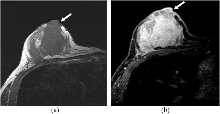
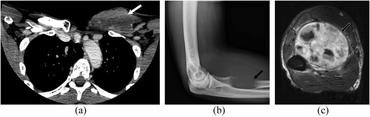
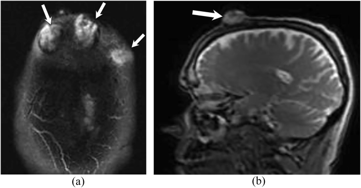
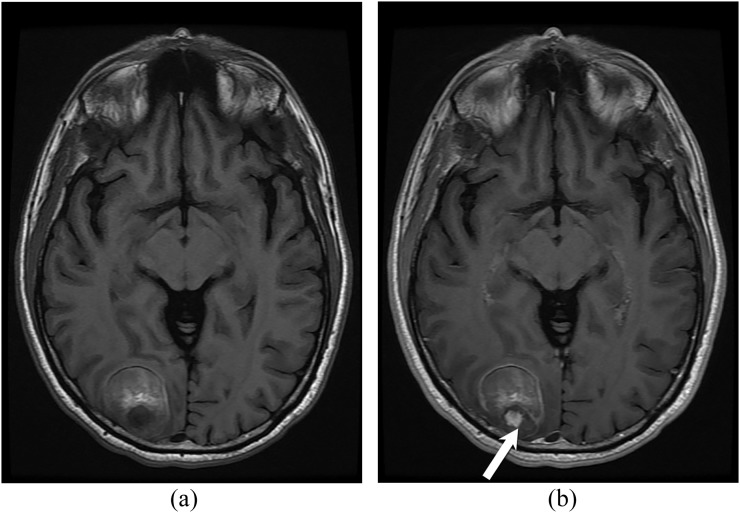
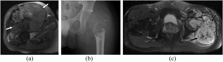
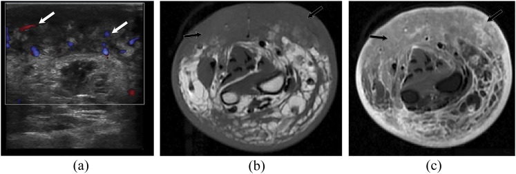
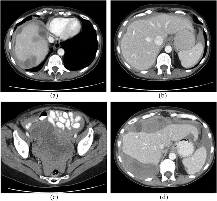
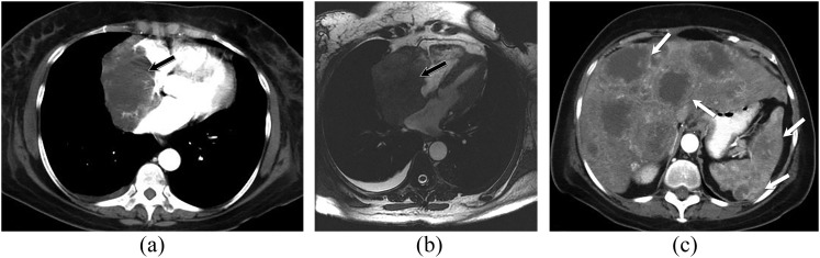
Angiosarcoma is a rare, aggressive subtype of soft-tissue sarcoma with a propensity for local recurrence and metastasis associated with a generally poor prognosis, unless diagnosed early. Given the vascular endothelial cell origin of angiosarcoma, tumours may develop in essentially any organ; however, there is a predilection for the skin where half of all tumours arise, increasing in prevalence with age. The most common risk factors are chronic lymphoedema and history of radiation. We review the most important radiological findings along the spectrum of angiosarcoma from head to toe throughout the body, including uncommon and rare locations. Key imaging features of angiosarcoma across multiple organ systems will be described, as well as the impact on management and prognosis.


Figures

Figure 1.
Figure 1.
A 43-year-old female with unilateral breast angiosarcoma. Axial T1 weighted images (a) before contrast and (b) after intravenous contrast administration showing a large irregular hypointense heterogeneously enhancing mass that near completely replaces the right breast. The mass extends to appear to involve the nipple/areolar complex (arrows).
Figure 2.
Figure 2.
A 47-year-old male with left chest wall angiosarcoma and left forearm metastases. Contrast-enhanced axial CT (a) image of the upper chest shows a large heterogeneous soft-tissue mass of the left anterior chest wall (white arrow). Forearm radiograph (b) demonstrates a lucent lytic lesion of the left proximal radius with soft-tissue swelling (black arrow). Axial T1 weighted MR image of the left forearm with intravenous contrast (c) shows an associated large heterogeneous enhancing soft-tissue mass of the flexor compartment (black-and-white arrow).
Figure 3.
Figure 3.
A 60-year-old male with cutaneous angiosarcoma of bilateral forehead. Axial (a) and sagittal (b) T2 weighted MR images show multifocal high signal intensity lesions arising from the skin of the forehead bilaterally (arrows). The lesions have exophytic components with associated osseous destruction of the outer table of the skull. No intracranial masses or extension.
Figure 4.
Figure 4.
A 61-year-old male with biopsy-proven primary brain angiosarcoma. Axial T1 weighted images before (a) and after (b) intravenous contrast demonstrate a roughly well-defined lesion in the right occipital lobe. The lesion has heterogeneous signal intensity with hyperintense components due to the presence of blood degradation products. A small nodule of enhancement seen posteriorly (arrow).
Figure 5.
Figure 5.
Two different patients with soft-tissue (a) and osseous angiosarcomas (b, c). (a) Axial T2 weighted MR image in a 63-year-old male with right thigh soft-tissue angiosarcoma. It demonstrates a large, well-defined, lobulated, solid, heterogeneous mass involving multiple muscles in the anterior compartment of the right thigh (white arrows). No underlying bone abnormality. (b, c) Plain radiograph and axial T2 weighted MR image in a 29-year-old male with angiosarcoma of the left femur. The plain radiograph (b) of the left femur demonstrates a large, expansile lytic lesion centred in the left femoral neck with associated pathological fracture and soft-tissue swelling. The axial T2 weighted MR image of the hip (c) shows a large, heterogeneous expansile aggressive tumour of the left proximal femur with extensive intramedullary involvement and pathological fracture as well as extension into the surrounding soft tissues (black arrows).
Figure 6.
Figure 6.
A 69-year-old male with chronic lymphoedema-induced angiosarcoma of the right forearm. The patient has a history of radical mastectomy and chronic lymphoedema. (a) Ultrasound image with Doppler of the right forearm shows diffuse soft-tissue mass with heterogeneous echotexture and increased vascularity (white arrows). Axial T1 weighted MR images (b) without and (c) with intravenous contrast demonstrate diffuse soft-tissue oedema with diffuse, extensive, infiltrative cutaneous and subcutaneous multinodular soft-tissue tumour along the volar tissues of the right forearm with involvement of underlying fascia and musculature (black arrows).
Figure 7.
Figure 7.
A 67-year-old male patient with primary peritoneal angiosarcoma presenting with haemoperitoneum. Contrast-enhanced CT images at presentation (a–c) show a diffuse peritoneal-based process with multifocal serosal implants on the liver (a, b) and large-volume high-attenuation peritoneal fluid consistent with haemorrhage. CT follow-up 1 month later (d) shows rapid progression of the heterogeneous peritoneal-based masses.
Figure 8.
Figure 8.
A 68-year-old female with cardiac angiosarcoma, hepatic and splenic metastases. Contrast-enhanced axial CT chest image (a) and axial bright-blood (b) MR images showing large discrete, soft-tissue mass that extends from the right atrial pericardium into the right atrium (black arrows). The mass occupies most of the right atrial cavity. Axial CT image of the upper abdomen (c) demonstrates multiple hypodense, peripherally enhancing lesions in the liver and spleen, consistent with metastatic disease (white arrows).
Figure 9.
Figure 9.
A 74-year-old male with angiosarcoma of the pulmonary artery. Axial CT angiography demonstrates massive filling defects in the main pulmonary artery, right and proximal left pulmonary arteries, simulating large pulmonary thromboembolism. Nodular borders, infiltration and enhancement help in distinguishing the mass from a bland thrombus.
Figure 10.
Figure 10.
A 54-year-old female with primary hepatic angiosarcoma. T1 weighted post-contrast MR images during arterial (a), portal venous (b) and delayed (c) phases show a dominant mass at the medial liver dome demonstrating progressive nodular enhancement that is similar to blood pool and resembles a cavernous haemangioma. However, multiple additional lesions are present, with a somewhat more irregular pattern of progressive enhancement. Similar to other vascular tumours, the masses are predominately hyperechoic at ultrasound (d). Contrast-enhanced CT images several months later in portal venous (e) and delayed (f) phases show marked interval tumour growth but with haemangioma-like enhancement pattern retained.
Figure 11.
Figure 11.
A 64-year-old female presenting with hepatic angiosarcoma, over 50 years after exposure to thorium dioxide (Thorotrast®). Abdominal radiograph (a) shows dense, shrunken spleen and dense abdominal lymph nodes compatible with prior Thorotrast exposure. Contrast-enhanced CT in the arterial phase (b) shows the same splenic and nodal findings plus multifocal hypervascular liver lesions, demonstrating both nodular and ring-like enhancement patterns.
Figure 12.
Figure 12.
A 62-year-old male with splenic angiosarcoma. (a) Unenhanced and (b) contrast-enhanced CT images show marked splenomegaly with irregular peripheral enhancement (b). Multiple liver lesions are also present, presumably from metastatic spread.
Figure 13.
Figure 13.
59- and 63-year-old males with penile and spermatic cord angiosarcomas. Contrast-enhanced axial CT images in two different patients with penile (a) and left spermatic cord angiosarcomas. There are heterogeneous diffusely infiltrative masses that involve the penis and encase the urethra in the first patient (a) as well as the left spermatic cord in the second patient (b) (arrows).

References

    1. Peterson MS, Baron RL, Rankin SC. Hepatic angiosarcoma: findings on multiphasic contrast-enhanced helical CT do not mimic hepatic hemangioma. AJR Am J Roentgenol 2000; 175: 165–70. doi: https://doi.org/10.2214/ajr.175.1.1750165 - DOI - PubMed
    1. Walker EA, Song AJ, Murphey MD. Magnetic resonance imaging of soft-tissue masses. Semin Roentgenol 2010; 45: 277–97. doi: https://doi.org/10.1053/j.ro.2009.12.004 - DOI - PubMed
    1. Young RJ, Brown NJ, Reed MW, Hughes D, Woll PJ. Angiosarcoma. Lancet Oncol 2010; 11: 983–91. doi: https://doi.org/10.1016/s1470-2045(10)70023-1 - DOI - PubMed
    1. Walker EA, Salesky JS, Fenton ME, Murphey MD. Magnetic resonance imaging of malignant soft tissue neoplasms in the adult. Radiol Clin North Am 2011; 49: 1219–34, vi. doi: https://doi.org/10.1016/j.rcl.2011.07.006 - DOI - PubMed
    1. Murinello A, Mendonca P, Abreu A, Santos AL, Roquete J, Pinto E, et al. . Cardiac angiosarcoma—a review. Rev Port Cardiol 2007; 26: 577–84. - PubMed