. 2017 May 4;18(5):973.
doi: 10.3390/ijms18050973.

Galectin-3 in Peripheral Artery Disease. Relationships with Markers of Oxidative Stress and Inflammation

Affiliations

Galectin-3 in Peripheral Artery Disease. Relationships with Markers of Oxidative Stress and Inflammation

Isabel Fort-Gallifa et al. Int J Mol Sci. .

Abstract

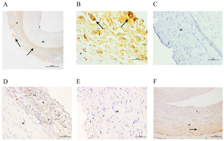
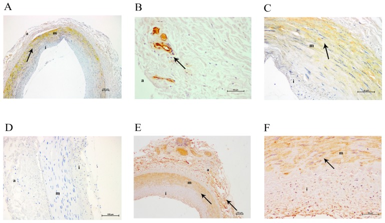
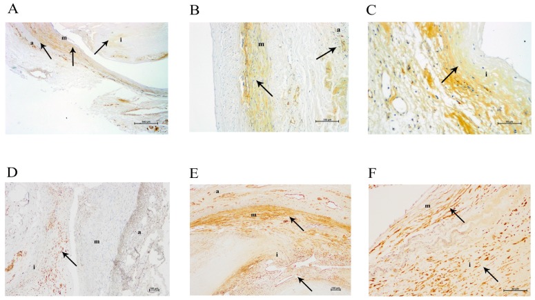
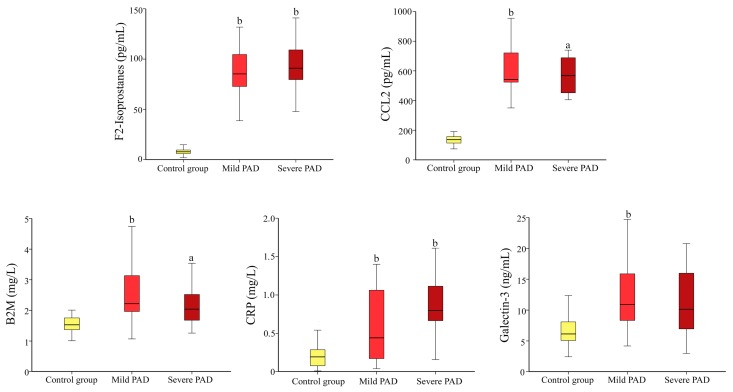
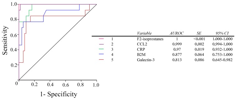
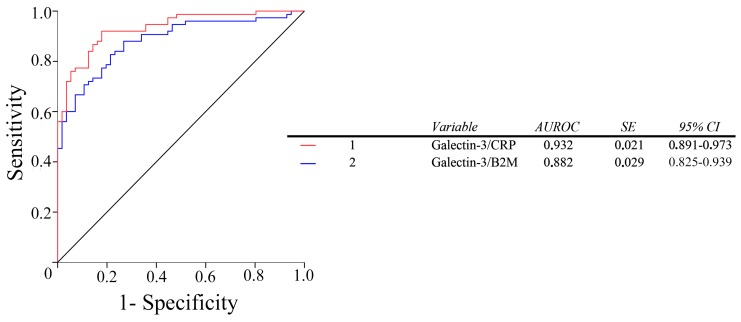
Galectin-3 is a modulator of oxidative stress, inflammation, and fibrogenesis involved in the pathogenesis of vascular diseases. The present study sought to characterize, in patients with peripheral artery disease (PAD), the localization of galectin-3 in arterial tissue, and to analyze the relationships between the circulating levels of galectin-3 and oxidative stress and inflammation. It also sought to compare the diagnostic accuracy of galectin-3 with that of other biochemical markers of this disease. We analyzed femoral or popliteal arteries from 50 PAD patients, and four control arteries. Plasma from 86 patients was compared with that from 72 control subjects. We observed differences in the expression of galectin-3 in normal arteries, and arteries from patients with PAD, with a displacement of the expression from the adventitia to the media, and the intima. In addition, plasma galectin-3 concentration was increased in PAD patients, and correlated with serologic markers of oxidative stress (F2-isoprostanes), and inflammation [chemokine (C-C motif) ligand 2, C-reactive protein, β-2-microglobulin]. We conclude that the determination of galectin-3 has good diagnostic accuracy in the assessment of PAD and compares well with other analytical parameters currently in use.

Keywords: F2-isoprostanes; atherosclerosis; galectin-3; oxidative stress; peripheral artery disease.


Conflict of interest statement

The authors declare no conflict of interest.

Figures

Figure 1
Figure 1
Immunohistochemical images of a normal femoral artery. (A) Galectin-3 staining in adventitia at a magnification of 40×; (B) galectin-3 staining in the adventitia at a magnification of 200×; (C) lack of galectin-3 staining in tunica media at a magnification of 200×; (D) faint CD68 staining in the adventitia at 200× magnification; (E) lack of CD68 staining in the media at 200× magnification; (F) α-actin staining in the media at 100× magnification. a, adventitia; m, media; i, intima. The arrows show positive immunostained areas.
Figure 2
Figure 2
Immunohistochemical images of a moderately altered femoral artery. (A) Galectin-3 staining at 40× magnification; (B) galectin-3 staining in adventitia at 200× magnification; (C) galectin-3 staining in tunica media at 200× magnification; (D) lack of CD68 staining at 100× magnification; (E) α-actin staining at 40× magnification; (F) α-actin staining at 200× magnification. a, adventitia; m, media; i, intima. The arrows show positive immunostained areas.
Figure 3
Figure 3
Immunohistochemical images of a severely altered femoral artery. (A) Galectin-3 staining at 40× magnification; (B) galectin-3 staining in adventitia at 100× magnification; (C) galectin-3 staining in tunica intima at 200× magnification; (D) CD68 staining at 100×magnification; (E) α-actin staining in adventitia, media, and intima at 100× magnification; (F) α-actin staining in media and intima at 200× magnification. a, adventitia; m, media; i, intima. The arrows show positive immunostained areas.
Figure 4
Figure 4
Selected biochemical variables in PAD patients classified according to whether they had mild disease (Fontaine Stages I and II) severe disease (Fontaine Stages III and IV), or were in the control group. Significance values by the Mann–Whitney U test: a p < 0.01; b p < 0.001, with respect to the control group.
Figure 5
Figure 5
Receiver operating characteristics (ROC) plots for all the studied biomarkers in PAD patients and in the control group. AUROC: areas under the curve of the ROC plots. SE: Standard Error.
Figure 6
Figure 6
Receiver operating characteristics (ROC) plots for the galectin-3/CRP and galectin-3/B2M ratios in PAD patients and in the control group. AUROC: areas under the curve of the ROC plots. SE: Standard Error.

References

    1. Barondes S.H., Cooper D.N., Gitt M.A., Leffler H. Galectins. Structure and function of a large family of animal lectins. J. Biol. Chem. 1994;269:20807–20810. - PubMed
    1. Pugliese G., Iacobini C., Pesce C.M., Menini S. Galectin-3: An emerging all-out player in metabolic disorders and their complications. Glycobiology. 2015;25:136–150. doi: 10.1093/glycob/cwu111. - DOI - PubMed
    1. Rubinstein N., Ilarregui J.M., Toscano M.A., Rabinovich G.A. The role of galectins in the initiation, amplification and resolution of the inflammatory response. Tissue Antigens. 2004;64:1–12. doi: 10.1111/j.0001-2815.2004.00278.x. - DOI - PubMed
    1. Paz I., Sachse M., Dupont N., Mounier J., Cederfur C., Enninga J., Leffler H., Poirier F., Prevost M.C., Lafont F., et al. Galectin-3, a marker for vacuole lysis by invasive pathogens. Cell. Microbiol. 2010;12:530–544. doi: 10.1111/j.1462-5822.2009.01415.x. - DOI - PubMed
    1. DeRoo E.P., Wrobleski S.K., Shea E.M., Al-Khalil R.K., Hawley A.E., Henke P.K., Myers D.D., Jr., Wakefield T.W., Diaz J.A. The role of galectin-3 and galectin-3-binding protein in venous thrombosis. Blood. 2015;125:1813–1821. doi: 10.1182/blood-2014-04-569939. - DOI - PMC - PubMed