Obestatin Plays Beneficial Role in Cardiomyocyte Injury Induced by Ischemia-Reperfusion In Vivo and In Vitro
- PMID: 28472020
- PMCID: PMC5426386
- DOI: 10.12659/msm.901361
Obestatin Plays Beneficial Role in Cardiomyocyte Injury Induced by Ischemia-Reperfusion In Vivo and In Vitro
Retraction in
-
Retracted: Obestatin Plays Beneficial Role in Cardiomyocyte Injury Induced by Ischemia-Reperfusion In Vivo and In Vitro.Med Sci Monit. 2023 Oct 4;29:e942752. doi: 10.12659/MSM.942752. Med Sci Monit. 2023. PMID: 37791420 Free PMC article.
Abstract
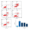
BACKGROUND Obestatin, primarily recognized as a peptide within the gastrointestinal system, has been shown to benefit the cardiovascular system. We designed this experiment to study the protective role and underlying mechanism of obestatin against ischemia-reperfusion(I/R) injury in myocardial cells. MATERIAL AND METHODS In an In vivo experiment, LAD was ligated for 0.5 h and then opened for reperfusion with obestatin for 24 h. Then, the infarction area was shown with TTC staining, and inflammation factors in serum were analyzed by qRT-PCR. In primary cultured cardiomyocytes, we measured the level of LDH, MDA, GSH, and SOD. Finally, we assessed cells apoptosis using flow cytometry and detected the concentrations of caspase-3, Bax, and Bcl-2 using Western blot analysis. RESULTS TTC staining showed that in the 3 obestatin groups, the infarct area became smaller with the increase of obestatin concentration. Obestatin also inhibited LDH expression in rat serum and decreased mRNA levels of TNF-α, IL-6, ICAM-1, and iNOS in rat cardiomyocytes after reperfusion. In primary cultured cardiomyocytes, obestatin decreased LDH content and increased GSH level after I/R injury. Obestatin was also found to antagonize the apoptosis of cardiomyocytes in a dose-dependent manner. Western blot analysis showed that obestatin downregulated the expression of caspase-3 and Bax and upregulated the expression of Bcl-2. CONCLUSIONS Obestatin can protect cardiomyocyte from I/R-induced injury in vitro and in vivo. This beneficial effect is closely related with its properties of anti-inflammation, anti-cytotoxicity, and anti-apoptosis. The protective effect of obestatin might be associated with activation of Bcl-2 and inhibition of caspase-3 and Bax.
Conflict of interest statement
The authors declare no financial conflicts of interest.
Figures


Similar articles
-
Influences of remifentanil on myocardial ischemia-reperfusion injury and the expressions of Bax and Bcl-2 in rats.Eur Rev Med Pharmacol Sci. 2018 Dec;22(24):8951-8960. doi: 10.26355/eurrev_201812_16665. Eur Rev Med Pharmacol Sci. 2018. PMID: 30575939
-
Cardioprotective effect of breviscapine: inhibition of apoptosis in H9c2 cardiomyocytes via the PI3K/Akt/eNOS pathway following simulated ischemia/reperfusion injury.Pharmazie. 2015 Sep;70(9):593-7. Pharmazie. 2015. PMID: 26492644
-
Resveratrol attenuates ischemia/reperfusion injury in neonatal cardiomyocytes and its underlying mechanism.PLoS One. 2012;7(12):e51223. doi: 10.1371/journal.pone.0051223. Epub 2012 Dec 20. PLoS One. 2012. PMID: 23284668 Free PMC article.
-
Obestatin affords cardioprotection to the ischemic-reperfused isolated rat heart and inhibits apoptosis in cultures of similarly stressed cardiomyocytes.Am J Physiol Heart Circ Physiol. 2010 Aug;299(2):H470-81. doi: 10.1152/ajpheart.00800.2009. Epub 2010 Jun 4. Am J Physiol Heart Circ Physiol. 2010. PMID: 20525876
-
Obestatin as a key regulator of metabolism and cardiovascular function with emerging therapeutic potential for diabetes.Br J Pharmacol. 2016 Jul;173(14):2165-81. doi: 10.1111/bph.13502. Epub 2016 May 27. Br J Pharmacol. 2016. PMID: 27111465 Free PMC article. Review.
Cited by
-
Therapeutic Peptides to Treat Myocardial Ischemia-Reperfusion Injury.Front Cardiovasc Med. 2022 Feb 17;9:792885. doi: 10.3389/fcvm.2022.792885. eCollection 2022. Front Cardiovasc Med. 2022. PMID: 35252383 Free PMC article. Review.
-
Potential Therapeutic Effects of Gut Hormones, Ghrelin and Obestatin in Oral Mucositis.Int J Mol Sci. 2019 Mar 27;20(7):1534. doi: 10.3390/ijms20071534. Int J Mol Sci. 2019. PMID: 30934722 Free PMC article. Review.
-
Investigation of the association between cardio-metabolic risk factors, neurotrophins and gastric hormones among apparently healthy women: A cross-sectional analysis.J Cardiovasc Thorac Res. 2022;14(1):53-60. doi: 10.34172/jcvtr.2022.10. Epub 2022 Mar 6. J Cardiovasc Thorac Res. 2022. PMID: 35620753 Free PMC article.
-
Long noncoding RNA-MEG3 contributes to myocardial ischemia-reperfusion injury through suppression of miR-7-5p expression.Biosci Rep. 2019 Aug 19;39(8):BSR20190210. doi: 10.1042/BSR20190210. Print 2019 Aug 30. Biosci Rep. 2019. PMID: 31366567 Free PMC article.
-
Small intestinal development in suckling rats after enteral obestatin administration.PLoS One. 2018 Oct 19;13(10):e0205994. doi: 10.1371/journal.pone.0205994. eCollection 2018. PLoS One. 2018. PMID: 30339696 Free PMC article.
References
-
- Zhao CM, Furnes MW, Stenström B, et al. Characterization of obestatin- and ghrelin-producing cells in the gastrointestinal tract and pancreas of rats: An immunohistochemical and electron-microscopic study. Cell Tissue Res. 2008;331(3):575–87. - PubMed
-
- Zhang JV, Ren PG, Avsian-Kretchmer O, et al. Obestatin, a peptide encoded by the ghrelin gene, opposes ghrelin’s effects on food intake. Science. 2005;310(5750):996–99. - PubMed
-
- Granata R, Gallo D, Luque RM, et al. Obestatin regulates adipocyte function and protects against diet-induced insulin resistance and inflammation. FASEB J. 2012;26(8):3393–411. - PubMed
Publication types
MeSH terms
Substances
LinkOut - more resources
Full Text Sources
Research Materials
Miscellaneous

