Review
. 2017 Jun 1;38(3):255-266.
doi: 10.1210/er.2017-00064.

Energy Metabolism of the Osteoblast: Implications for Osteoporosis

Affiliations
Review

Energy Metabolism of the Osteoblast: Implications for Osteoporosis

Wen-Chih Lee et al. Endocr Rev. .

Abstract

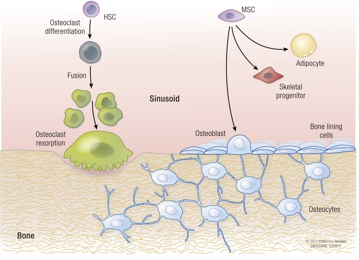
Osteoblasts, the bone-forming cells of the remodeling unit, are essential for growth and maintenance of the skeleton. Clinical disorders of substrate availability (e.g., diabetes mellitus, anorexia nervosa, and aging) cause osteoblast dysfunction, ultimately leading to skeletal fragility and osteoporotic fractures. Conversely, anabolic treatments for osteoporosis enhance the work of the osteoblast by altering osteoblast metabolism. Emerging evidence supports glycolysis as the major metabolic pathway to meet ATP demand during osteoblast differentiation. Glut1 and Glut3 are the principal transporters of glucose in osteoblasts, although Glut4 has also been implicated. Wnt signaling induces osteoblast differentiation and activates glycolysis through mammalian target of rapamycin, whereas parathyroid hormone stimulates glycolysis through induction of insulin-like growth factor-I. Glutamine is an alternate fuel source for osteogenesis via the tricarboxylic acid cycle, and fatty acids can be metabolized to generate ATP via oxidative phosphorylation although temporal specificity has not been established. More studies with new model systems are needed to fully understand how the osteoblast utilizes fuel substrates in health and disease and how that impacts metabolic bone diseases.


Figures

Figure 1.
Figure 1.
The bone remodeling unit is composed of several distinct cell types that originate from either the hematopoietic or mesenchymal lineages. Osteocytes are the “command and control” cells that regulate both resorption by osteoclasts and formation by osteoblasts. Bone-lining cells and mesenchymal stromal cells can differentiate into osteoblasts or adipocytes. Osteoblast progenitor differentiation is very dependent on glycolytic pathways. HSC, hematopoietic stem cell; MSC, mesenchymal stromal cell.
Figure 2.
Figure 2.
Metabolic fates of glucose in mammalian cells. Major biochemical pathways are denoted in red, with the main product from each pathway shown in blue. Glucose is used to produce not only energy but also intermediate metabolites for biosynthesis. Several key enzymes are highlighted in green. Note that citrate can be exported from mitochondria and converted to acetyl-coA in the nucleus to exert epigenetic regulation on gene expression. Although the metabolic pathways are common among different cell types, their relative importance likely varies depending on the biological function of each cell. G3pdh, glycerol-3-phosphate dehydrogenase; G6pdh, G6P dehydrogenase; Gfat, glutamine–fructose-6-phosphate transaminase; Pdh, pyruvate dehydrogenase; Phgdh, phosphoglycerate dehydrogenase; Ldha, lactate dehydrogenase; OAA, oxaloacetate; PRPP, phosphoribosyl pyrophosphate; UDPGlcNAc, uridine diphosphate N-acetylglucosamine.
Figure 3.
Figure 3.
Glutamine plays multiple roles in mammalian cells. Major biochemical pathways are shown in red. Glutamine can serve as a major energy source through oxidative phosphorylation in mitochondria. This process requires the conversion of glutamine to α-ketoglutarate, which is controlled by several key enzymes highlighted in green. Alt, alanine transaminase; Ast, aspartate transaminase; Gdh, glutamate dehydrogenase; Gls, glutaminase.
Figure 4.
Figure 4.
Wnt signaling stimulates aerobic glycolysis, glutamine catabolism, and fatty acid oxidation in osteoblast-lineage cells. Wnt-mTOR signaling acutely increases the protein but not mRNA levels of key enzymes involved in glucose and glutamine metabolism. Wnt also signals through β-catenin to increase the mRNA levels of genes important for fatty acid oxidation. Acad, acyl-coA dehydrogenase; Cpt1, carnitine palmitoyl transferase 1; Fz, frizzled; Gls, glutaminase; Hadha, hydroxyacyl–coenzyme A dehydrogenase/3-ketoacyl–coenzyme A thiolase/enoyl–coenzyme A hydratase (trifunctional protein), α subunit; Hk2, hexokinase 2; Ldha, lactate dehydrogenase A; Pdc, pyruvate dehydrogenase; Pfk1, phosphofructokinase 1.

References

    1. Borle AB, Nichols N, Nichols G Jr. Metabolic studies of bone in vitro. I. Normal bone. J Biol Chem. 1960;235:1206–1210. - PubMed
    1. Cohn DV, Forscher BK. Aerobic metabolism of glucose by bone. J Biol Chem. 1962;237:615–618. - PubMed
    1. Peck WA, Birge SJ Jr, Fedak SA. Bone Cells: Biochemical and Biological Studies after Enzymatic Isolation. Science. 1964;146(3650):1476–1477. - PubMed
    1. Peck WA, Birge SJ Jr, Brandt J. Collagen synthesis by isolated bone cells: stimulation by ascorbic acid in vitro. Biochim Biophys Acta. 1967;142(2):512–525. - PubMed
    1. Gerstenfeld LC, Chipman SD, Glowacki J, Lian JB. Expression of differentiated function by mineralizing cultures of chicken osteoblasts. Dev Biol. 1987;122(1):49–60. - PubMed

Publication types

Substances