. 2017 Jun 20;45(11):6894-6910.
doi: 10.1093/nar/gkx307.

WIG1 is crucial for AGO2-mediated ACOT7 mRNA silencing via miRNA-dependent and -independent mechanisms

Affiliations

WIG1 is crucial for AGO2-mediated ACOT7 mRNA silencing via miRNA-dependent and -independent mechanisms

Hyung Chul Lee et al. Nucleic Acids Res. .

Abstract

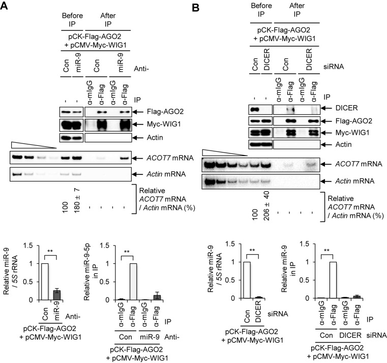
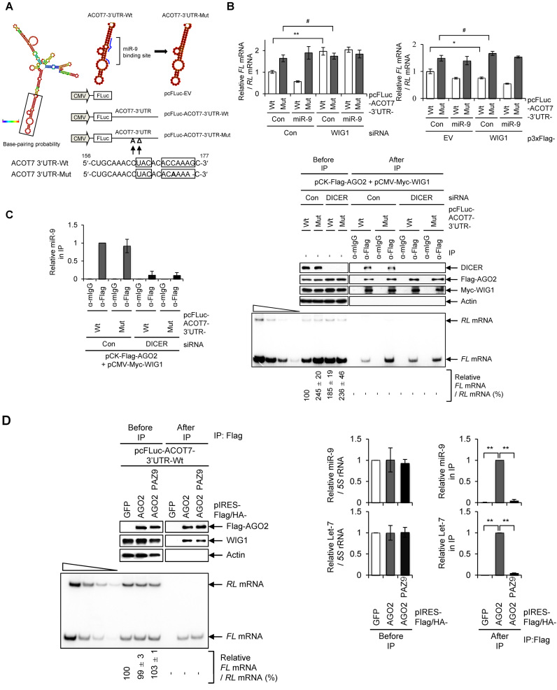
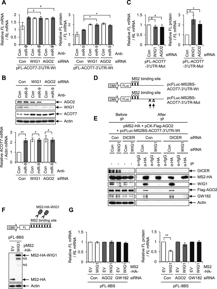
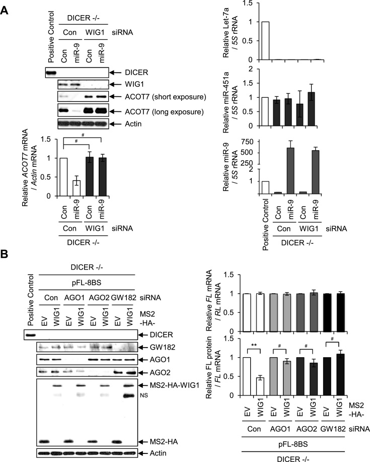
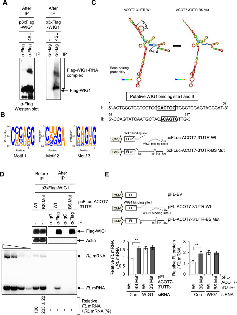
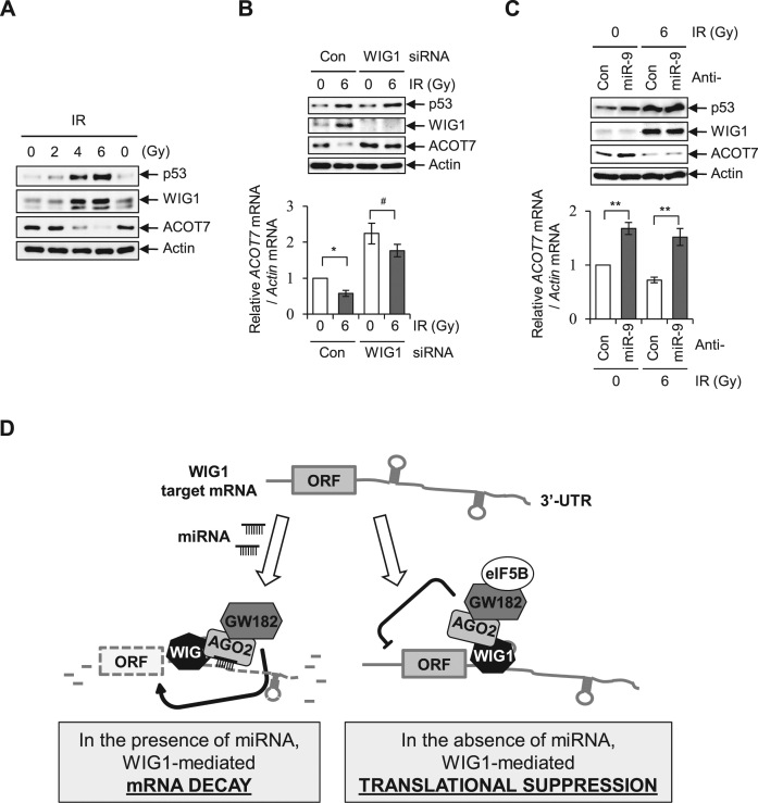
RNA-binding proteins (RBPs) are involved in mRNA splicing, maturation, transport, translation, storage and turnover. Here, we identified ACOT7 mRNA as a novel target of human WIG1. ACOT7 mRNA decay was triggered by the microRNA miR-9 in a WIG1-dependent manner via classic recruitment of Argonaute 2 (AGO2). Interestingly, AGO2 was also recruited to ACOT7 mRNA in a WIG1-dependent manner in the absence of miR-9, which indicates an alternative model whereby WIG1 controls AGO2-mediated gene silencing. The WIG1-AGO2 complex attenuated translation initiation via an interaction with translation initiation factor 5B (eIF5B). These results were confirmed using a WIG1 tethering system based on the MS2 bacteriophage coat protein and a reporter construct containing an MS2-binding site, and by immunoprecipitation of WIG1 and detection of WIG1-associated proteins using liquid chromatography-tandem mass spectrometry. We also identified WIG1-binding motifs using photoactivatable ribonucleoside-enhanced crosslinking and immunoprecipitation analyses. Altogether, our data indicate that WIG1 governs the miRNA-dependent and the miRNA-independent recruitment of AGO2 to lower the stability of and suppress the translation of ACOT7 mRNA.


Figures

Figure 1.
Figure 1.
WIG1 regulates ACOT7 mRNA levels via miR-9-mediated mRNA decay. (A) Association of WIG1 with ACOT7 mRNA. MCF7 cells were harvested 2 days after transfection with p3xFlag-WIG1, and RIP analysis was performed using an anti-Flag M2 affinity gel. Immunoprecipitated proteins were subjected to immunoblotting using an anti-Flag antibody (top); RNAs were radiolabeled by RT-PCR using ACOT7-specific primers (bottom), and band intensities were quantified using Quantity One software. The four leftmost lanes represent 2-fold serial dilutions of RNA and confirm that RT-PCR was semi-quantitative. Data are expressed as the means ± SD from three independent experiments. (B) Effects of WIG1 depletion on ACOT7 mRNA and protein levels. MCF7 cells were transfected with WIG1 Si, and qRT-PCR and western blotting analyses were performed. Ectopic expression of WIG1 downregulates ACOT7 mRNA and protein levels. MCF7 cells transfected with p3xFlag-WIG1 construct were harvested and analyzed as described in the left panel. (C) Binding of AGO2 to ACOT7 mRNA in a WIG1-dependent manner. HEK 293T cells were harvested 2 days after transfection with either WIG1 or AGO2 Si, after which an RNP-IP assay was performed using anti-AGO2 or anti-WIG1 antibody, respectively. Immunoprecipitated proteins were subjected to immunoblotting using anti-AGO2 and anti-WIG1 antibody (left). Isolated RNAs were performed with qRT-PCR for quantification of ACOT7 mRNA levels (right). Actin mRNA and protein were used as a negative or normalized control. (D) Effects of miR-9 on ACOT7 mRNA and ACOT7 protein levels in WIG1-depleted MCF7 cells. Cell lysates were prepared 2 days after transfection of miR-9 into WIG1 Si-transfected cells and subjected to western blot analysis (top). RNAs were isolated, and RT-qPCR analysis was used to quantify ACOT7 mRNA levels (bottom). (E) Effects of miR-9 on ACOT7 mRNA and protein levels in WIG1-overexpressing MCF7 cells. p3xFlag-WIG1 plasmids were transfected into miR-9-transfected cells; 2 days later, protein levels were assessed by immunoblot analysis (top) and ACOT7 mRNA levels by RT-qPCR analysis (bottom). (F) WIG1-dependent binding of miR-9 to ACOT7 mRNA. Cell lysates were harvested and pulled-down with streptavidin beads 2 days after transfection with biotin-miR-9 in WIG1-depleted HEK 293T cells. Pulled-down RNA was amplified by RT-sqPCR using ACOT7-specific primers and quantified. Actin mRNA levels served as internal controls. The four leftmost lanes represent sequential 2-fold dilutions of RNA. Data are expressed as the means ± SD from three independent experiments. (G) Presence of miR-9 on target ACOT7 mRNA under WIG1-overexpressing conditions. Cell lysates were harvested and precipitated with streptavidin beads 2 days after transfection with biotin-miR-9 in WIG1-overexpressing HEK 293T cells. Precipitated RNAs were analyzed as described in panel F. *P < 0.05; **P < 0.01; #P > 0.05.
Figure 2.
Figure 2.
Inhibition of miRNA processing and disruption of ACOT7-3΄UTR miR-9-binding site are not sufficient to block AGO2-WIG1 binding to ACOT7 mRNA. (A) Effect of anti-miR-9-5p on AGO2-WIG1 binding to ACOT7 mRNA. HEK 293T cell lysates were harvested and precipitated with anti-Flag antibody 2 days after co-transfection of pCK-Flag-AGO2 and pCMV-Myc-WIG1 plasmids in combination with anti-miR-9. From RNP-IP, precipitated proteins were subjected to Western blotting (top). RNAs in precipitates were radiolabeled with RT-PCR using ACOT7-specific primers (bottom). Actin mRNA served as a negative control. Band intensity was quantified using Quantity One software. The four leftmost lanes represent 2-fold serial dilutions of RNA, and confirm RT-PCR was semi-quantitative. Relative level of miR-9 in anti-miR-9-treated cells was normalized to that of a 5S rRNA internal control, relative to anti-miR-Con. Fold enrichment of miR-9 in Flag-AGO2 immunoprecipitates was assessed after treatment with anti-miR-9, relative to anti-miR-Con. Data are presented as means ± SD from three independent experiments. (B) Binding of AGO2-WIG1 to ACOT7 mRNA in DICER-silenced cells. Immunoprecipitation was performed with anti-Flag antibody 2 days after co-transfection of pCK-Flag-AGO2 and pCMV-Myc-WIG1 in DICER Si-transfected HEK 293T cells. Precipitated proteins were subjected to western blotting (top). RNAs in precipitates were subjected to RT-PCR using ACOT7 mRNA-specific primers (bottom). Relative level of miR-9 in DICER-treated cells was normalized to that of a 5S rRNA internal control, relative to anti-miR-Con. Fold enrichment of miR-9 in Flag-AGO2 immunoprecipitates was assessed after treatment with DICER Si, relative to anti-miR-Con. Data are presented as means ± SD from three independent experiments. *P < 0.05; **P < 0.01; #P > 0.05.
Figure 3.
Figure 3.
WIG1 binds to ACOT7 mRNA 3΄UTR and recruits AGO2 despite disruption of miR-9 site, inhibition of miRNA synthesis or loss of miRNA. (A) Cloning strategy for disrupting miR-9 target sequence on ACOT7 3‘UTR with stem loops as predicted by RNAfold software. ACOT7 3΄UTR containing one substitution (172C → A) and one deletion (176G → Δ) in miR-9 target site was cloned into pcFLuc-empty vector (EV); Wt, wild-type; Mut, mutant. pcFLuc-EV was used as control. Base-pair probability is color-coded. (B) Effect of miR-9 on ACOT7-3΄UTR-Wt and -Mut reporters after modulation of WIG1 levels. Cells were transfected with either WIG1 Si or p3xFlag-WIG1 (Flag-Wig), followed by either pcFLuc-ACOT7-3΄UTR-Wt or -Mut in combination with miR-9. pRL-CMV was used as a reference plasmid. RNAs were isolated 2 days after transfection and measured by RT-qPCR analysis. FL mRNA levels were normalized to those expressed from pcFLuc-EV mRNA; RT-PCR intensities represent the means ± SD from three independent experiments. (C) Binding of AGO2 to ACOT7 3΄UTR containing mutations in miR-9 target sites. HEK 293T cells were transfected with DICER Si and then transfected with either pcFLuc-ACOT7-3΄UTR-Wt or -Mut reporter plasmids, pCK-Flag-AGO2 and pCMV-Myc-WIG1 in combination with pRL-CMV reference plasmid. Left, following RIP, precipitated proteins were assessed by western blot analysis to identify the indicated proteins (top), RNAs were radiolabeled using RT-PCR with FL and RL mRNA-specific primers and band intensities were quantified using Quantity One software (bottom). The four leftmost lanes represent sequential 2-fold dilutions of RNA. Left, miR-9 levels in Flag-AGO2 immunoprecipitates, as assessed by RT-qPCR analysis using pcFLuc-ACOT7-3΄UTR-Wt and -Mut reporters. Data represent the means ± SD from three independent experiments. (D) MicroRNA binding-defective AGO2 PAZ9 mutant binds to ACOT7 mRNA in the presence of WIG1. HEK 293T cells were transfected with pcFLuc-ACOT7-3΄UTR-Wt in combination with either pIRES-Flag/HA-AGO2 or -AGO2 PAZ9, after which RIP was performed using anti-Flag antibody. Precipitated proteins were assessed by western blot analysis (top), and precipitated RNAs were assessed by RT-qPCR analysis (bottom). pRL-CMV was used as a negative control for IP efficiency. Levels of miRNAs extracted from pre-immune lysates (Before IP) or immunoprecipitates (After IP) were assessed by RT-qPCR (right). Data represent the means ± SD from three independent experiments. *P < 0.05; **P < 0.01; #P > 0.05.
Figure 4.
Figure 4.
WIG1 tethering suppresses ACOT7 protein synthesis in AGO2-GW182 dependency. (A) Relative translational efficiency of ACOT7 3΄UTR reporters in the presence of anti-miR-9 in WIG1- or AGO2-depleted cells. MCF7 cells were transfected with either WIG1 Si or AGO2 Si as well as with pFL-ACOT7-3΄UTR-Wt and anti-miR-9 in combination with reference plasmid pRL-CMV. Cells were harvested 2 days after transfection, and FL and RL mRNAs were quantified using RT-qPCR. FL mRNA levels were normalized to RL mRNA levels (left). Translation rates were determined as the ratios of FL protein (luciferase activity, normalized to RL) to FL mRNA (normalized to RL mRNA). (B) Endogenous ACOT7 mRNA and protein levels in the presence of anti-miR-9 in WIG1- or AGO2-depleted cells. Lysates were prepared as explained in Figure 4A, followed by western blot analysis. Total RNA was subjected by qRT-PCR using ACOT7-specific primers. Actin was used as a loading control. (C) MCF7 cells were transfected with either WIG1 Si or AGO2 Si in combination with pFL-ACOT7-3΄UTR-Mut and pRL-CMV reference plasmid. FL mRNA levels and translation rates were determined as explained in panel A. (D) Diagrams of reporter vectors encoding ACOT7 mRNA 3΄UTR-Wt and -Mut with three copies of MS2-binding site (BS): pcFLuc-MS2BS-ACOT7-3΄UTR-Wt and -Mut. pcFLuc-EV was used as a reference control. (E) WIG1-dependent recruitment of AGO2-GW182 to 3΄UTR of ACOT7 mRNA in the absence of miRNA processing. HEK 293T cells were transfected with DICER Si and WIG1 Si and pcFLuc-MS2BS-ACOT7-3΄UTR-Wt reporter plasmid in combination with pCK-Flag-AGO2 and pMS2-HA. Cells were harvested 2 days after transfection and subjected to IP and western blot analysis to detect the indicated proteins. (F) Diagrams of MS2-HA-WIG1 reporter vector encoding FL coding region with eight copies of MS2-binding site (BS). (G) MCF7 cells were transfected either AGO2 Si or GW182 Si, followed by pFL-8BS, pMS2-HA and pMS2-HA-WIG1 in combination with pRL-CMV reference plasmid. Cells were harvested 2 days after transfection, followed by western blot analysis. Reporter RNA levels and translation rates were determined as explained in panel A. Data are presented as means ± SD from three independent experiments. *P < 0.05; **P < 0.01; #P > 0.05.
Figure 5.
Figure 5.
Effects of WIG1 on ACOT7 mRNA and protein levels in human HCT116 DICER knockout (−/−) cell line. (A) WIG depletion induces upregulation of ACOT7 protein levels without alteration of mRNA levels, regardless of miR-9, in DICER −/− cells. Cells were harvested at 2 days after miR-9 transfection and subjected to western blot analysis. RT-qPCR analysis was used to quantify specific RNA levels. Let-7a and miR-451a were used as positive and negative controls for DICER-dependent miRNA-maturation products, respectively. (B) WIG1 tethering in the absence of AGO1, AGO2 or GW182 induces translational suppression in DICER −/− cells. pFL-8BS reporters in combination with pMS2-HA plasmids were transfected in AGO1, AGO2 or GW182 Si-transfected DICER −/− cells. Two days after transfection, cell lysates analyzed with western blotting and RT-qPCR. Translation rate was measured as the ratio of FL protein to mRNA. HCT116 parental cells were used as a positive control regarding DICER −/− cells. Data are represented as the means ± SD from three independent experiments. **P < 0.01; #P > 0.05.
Figure 6.
Figure 6.
WIG1 prevents eIF5B assembly during translational initiation of 60S ribosomal subunit joining via AGO2-GW182 recruitment under miRNA deficiency. (A) Schematic representations of IRES constructs encoding FL with ACOT7 mRNA 3΄UTR. pFL-EV was used as a normalization control. (B) WIG1 suppresses translational initiation in the ACOT7-3΄UTR. MCF7 cells were transfected with WIG1 Si and pFL-ACOT7-3΄UTR-Wt, pEMCV-FL-ACOT7-3΄UTR-Wt or pCrPV-FL-ACOT7-3΄UTR-Wt in combination with pRL-CMV reference plasmid. Cells were harvested 24 h after transfection, and FL mRNA and (normalization control) RL mRNA were quantified by RT-qPCR analysis (top). Normalized level of each IRES reporter mRNA in the presence of each Con Si was set as 1. Translation rates were calculated as explained in Figure 4A. FL protein levels (FL activity) were normalized to RL levels (RL activity) (bottom); normalized levels of FL/RL activity in the presence of Con Si were set as 1. (C) MiR-9 is not required for WIG1-mediated translational initiation of ACOT7 mRNA. MCF7 cells were transfected with WIG1 Si and pFL-ACOT7-3΄UTR-Mut, pEMCV-FL-ACOT7-3΄UTR-Mut or pCrPV-FL-ACOT7-3΄UTR-Mut in combination with pRL-CMV reference plasmid. Cells were harvested 24 h after transfection and FL mRNA and RL mRNA (for normalization) were quantified RT-qPCR (top). Normalized levels of each IRES reporter mRNA in the presence of each Con Si was set as 1. Translation rate was determined as the ratio of normalized FL protein to FL mRNA (as defined in Figure 4A); normalized level of FL activity in the presence of Con Si was defined as 1. Data are represented as the means ± SD from three independent experiments. **P < 0.01. (D) RNA-independent interaction between WIG1 and eIF5B. HEK 293T cell lysates were obtained from WIG1-overexpressing HEK 293T cells. After immunoprecipitation using anti-Flag M2 affinity gel, proteins in the IP material were detected by western blot analysis (top). RT-qPCR detection of Actin mRNA was performed to assess completion of RNase A digestion (bottom). (E) EIF5B was preferentially recruited to WIG1-target mRNAs and specifically associates with AGO2-GW182. After transfection of HEK 293T cells with either AGO2 Si or GW182 Si and subsequent transfection with p3xFlag-WIG1, anti-Flag was used for immunoprecipitation, and the indicated proteins were detected by immunoblotting.
Figure 7.
Figure 7.
Identification of WIG1-binding sequence by PAR-CLIP analysis. (A) 5’-end 32P-labeled RNA-Flag-WIG1 immunoprecipitates were prepared from RNase T1-treated lysates in the presence or absence of 100 mM 4-thiouridine (4SU) photoactivatable nucleoside and crosslinked with UV 365 nm (right). Western blot analysis using anti-Flag antibody (left). (B) Representative RNA recognition elements (RREs) of the top three significantly enriched motifs derived from motif analysis of WIG1 PAR-CLIP. (C) Cloning strategy for disruption of putative WIG1 PAR-CLIP site on the 3΄UTR of ACOT7 mRNA with secondary structure predicted by RNAfold software. ACOT7-3΄UTR-BS Mut lacking two putative WIG1-binding sites (BS) from the PAR-CLIP analysis was cloned into pcFLuc vector. Base-pairing probabilities are color-coded. (D) Deletion of putative WIG1-binding sites disrupts WIG1 binding to the ACOT7 3΄UTR. HEK 293T cells were transfected with either pcFLuc-ACOT7-3΄UTR-Wt or -BS-Mut in combination with p3xFlag-WIG1 and pRL-CMV as a reference plasmid. After RIP using anti-Flag antibody, western blot analysis was performed using anti-Flag antibody (top) and RL and FL mRNAs RNAs were analyzed by quantification of radiolabeled RT-PCR products using Quantity One software (bottom). The four leftmost lanes represent sequential 2-fold dilutions of RNA. pRL-CMV were used as a negative control. The relative ratio of RT-sqPCR intensity represents the mean ± SD from three independent experiments. (E) Relative translational efficiency of ACOT7-3΄UTR reporters in WIG1-depleted cells. Diagrams of reporter vectors encoding ACOT7 mRNA 3΄UTR-BS-Mut with full-length FL coding region. pFL-EV or pFL-ACOT7-3΄UTR-Wt was used as normalization control or positive control, respectively. Two days after transfection of MCF7 cells with WIG1 Si and pFL-ACOT7-3΄UTR reporters (pFL-EV, -Wt, -BS Mut) and pRL-CMV reference plasmid, RNA was isolated and FL mRNA and RL mRNA (normalization control) quantified by RT-qPCR analysis (left). Protein levels were determined with luciferase activity assay (right), and translation rates were determined as explained in Figure 4A. **P < 0.01.
Figure 8.
Figure 8.
Regulation of ACOT7 mRNA and protein levels in a WIG1-dependent manner following irradiation. (A) MCF7 cells were harvested after exposure to 2, 4 or 6 Gy of ionizing radiation (IR) and subjected to immunoblotting for detection of p53, WIG1 and ACOT7. (B) Cells were transfected with Con Si or WIG1 Si, exposed to 6 Gy of IR, incubated for 24 h and then harvested for immunoblot and RT-PCR analyses. (C) After transfection with anti-miR-9, MCF7 cells were exposed to IR and then harvested for immunoblot and RT-PCR analyses. Actin was used as an internal control (A–C). Each bar in the graphs represents the mean ± SD compared to control cells from three independent experiments (B and C). *P < 0.05; **P < 0.01; #P > 0.05. (D) Proposed model for novel dual role of WIG1: miRNA-dependent mRNA decay and miRNA-independent translational suppression. RNA-binding protein WIG1 is a novel dual repressor of gene silencing. WIG1 regulates target mRNA stability in an miRNA-dependent manner and suppresses translational initiation in an miRNA-independent manner.

Similar articles

Cited by

References

    1. Yi R., Qin Y., Macara I.G., Cullen B.R.. Exportin-5 mediates the nuclear export of pre-microRNAs and short hairpin RNAs. Genes Dev. 2003; 17:3011–3016. - PMC - PubMed
    1. Carthew R.W., Sontheimer E.J.. Origins and mechanisms of miRNAs and siRNAs. Cell. 2009; 136:642–655. - PMC - PubMed
    1. van Kouwenhove M., Kedde M., Agami R.. MicroRNA regulation by RNA-binding proteins and its implications for cancer. Nat. Rev. Cancer. 2011; 11:644–656. - PubMed
    1. Kim V.N., Han J., Siomi M.C.. Biogenesis of small RNAs in animals. Nat. Rev. Mol. Cell Biol. 2009; 10:126–139. - PubMed
    1. Bartel D.P. MicroRNAs: target recognition and regulatory functions. Cell. 2009; 136:215–233. - PMC - PubMed

MeSH terms