Review
. 2017 May 5;356(6337):eaaf3627.
doi: 10.1126/science.aaf3627.

Advances in engineering hydrogels

Affiliations
Review

Advances in engineering hydrogels

Yu Shrike Zhang et al. Science. .

Abstract

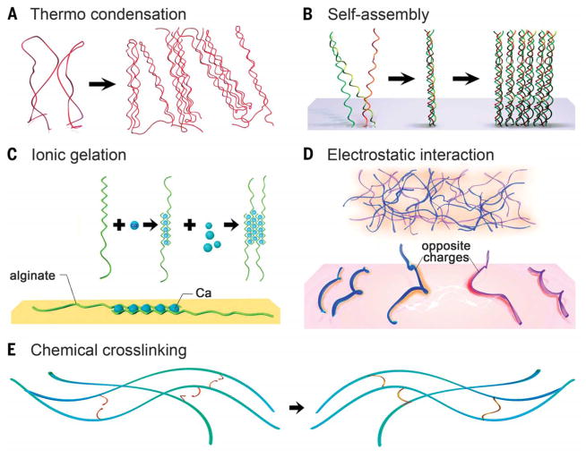
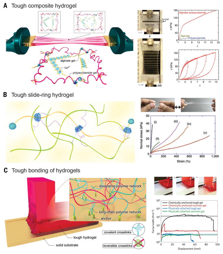
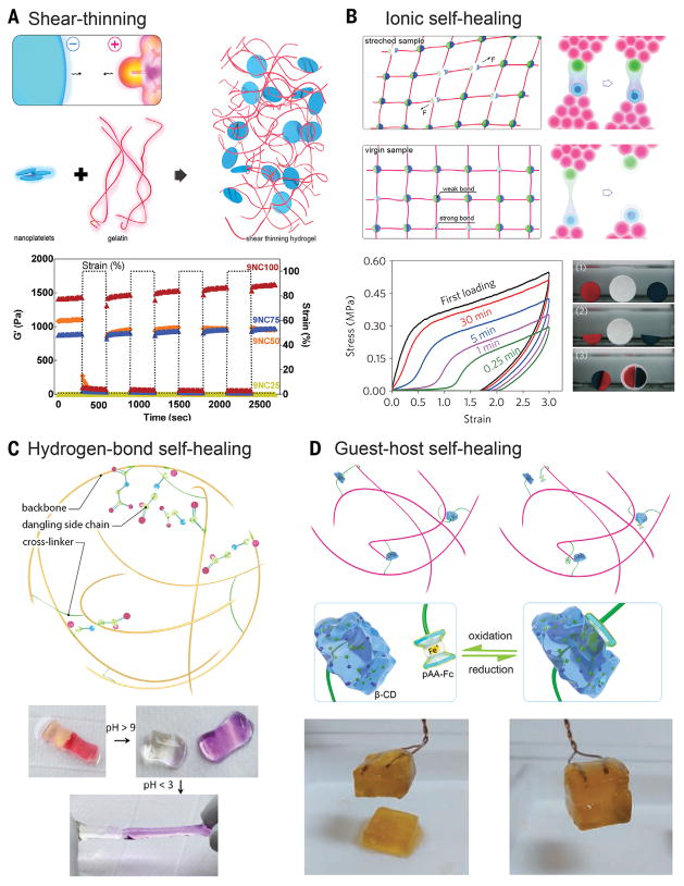
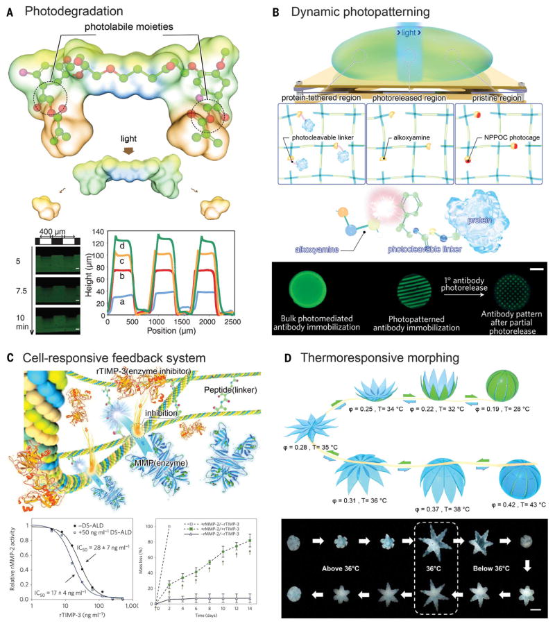
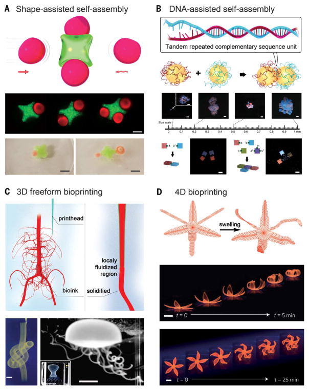
Hydrogels are formed from hydrophilic polymer chains surrounded by a water-rich environment. They have widespread applications in various fields such as biomedicine, soft electronics, sensors, and actuators. Conventional hydrogels usually possess limited mechanical strength and are prone to permanent breakage. Further, the lack of dynamic cues and structural complexity within the hydrogels has limited their functions. Recent developments include engineering hydrogels that possess improved physicochemical properties, ranging from designs of innovative chemistries and compositions to integration of dynamic modulation and sophisticated architectures. We review major advances in designing and engineering hydrogels and strategies targeting precise manipulation of their properties across multiple scales.


Figures

Fig. 1
Fig. 1. Cross-linking of hydrogels
(A to D) Physical cross-linking. (A) Thermally induced entanglement of polymer chains. (B) Molecular self-assembly. (C) Ionic gelation. (D) Electrostatic interaction. (E) Chemical cross-linking.
Fig. 2
Fig. 2. Tuning the mechanics of hydrogels
(A) Stretchable hydrogel made from long-chain polymers and reversible physically cross-linked polymers. (Right) The hydrogel could be stretched to 21 times its initial length, where the stretch λ is the final length of the unclamped region divided by the original length; stress-stretch curves of the alginate, PAAm, and alginate-PAAm hydrogels, each stretched to rupture, where the nominal stress s is defined as the force applied on the deformed hydrogel, divided by the cross-sectional area of the undeformed hydrogel. [Adapted with permission from (41), copyright 2012 Nature Publishing Group] (B) Stretchable hydrogel based on a sliding ring mechanism. (Right) Photo of an elongated NIPAAM-AAcNa-HPR-C hydrogel, and stress-strain curves of different hydrogels: (i) NIPAAM-AAcNa-BIS [0.65 weight % (wt %)], (ii) NIPAAM-AAcNa-BIS (0.065 wt %), (iii) NIPAAM-AAcNa-HPR-C (2.00 wt %), (iv) NIPAAM-AAcNa-HPR-C (1.21 wt %), and (v) NIPAAM-AAcNa-HPR-C (0.65 wt %). The percentages denote those of the cross-linkers. [Adapted with permission from (50), copyright 2014 Nature Publishing Group] (C) Tough bonding of hydrogels with smooth surfaces. (Right) The peeling process of a tough hydrogel chemically anchored on a glass substrate, and curves of the peeling force per width of hydrogel sheet versus displacement for various types of hydrogel-solid bonding. [Adapted with permission from (59), copyright 2016 Nature Publishing Group]
Fig. 3
Fig. 3. Shear-thinning and self-healing hydrogels
(A) Shear-thinning hydrogel through nanocompositing. (Bottom) Recovery of the nanocomposites was observed by subjecting the hydrogel to alternating high and low strain conditions (100% strain and 1% strain) while monitoring the moduli of the composite. [Adapted with permission from (60), copyright 2014 American Chemical Society] (B to D) Self-healing hydrogels based on ionic interactions, hydrogen bonds, and host-guest coupling. (B) Ionic interactions. (Bottom left) Recovery of the sample for different waiting times. (Bottom right) Self-healing between either two freshly cut surfaces (red and blue) or a fresh and an aged surface (white) of samples; [Adapted with permission from (55), copyright 2013 Nature Publishing Group] (C) Hydrogen bonds. (Bottom) The healed hydrogels at low pH separate after exposure to a high-pH solution (with pH > 9), and the separated hydrogels could reheal upon exposure to acidic solution (pH < 3). [Adapted with permission from (62), copyright 2012 the National Academy of Sciences] (D) Host-guest coupling. (Bottom) The cut hydrogel spread with NaClO aqueous solution did not heal after 24 hours, but readhesion was observed 24 hours after spreading reuced glutathione aqueous solution onto the oxidized cut surface. [Adapted with permission from (66), copyright 2011 Nature Publishing Group]
Fig. 4
Fig. 4. Dynamic modulation of hydrogel microenvironment
(A) Photo-degradation through photolabile moieties. (Bottom left) Hydrogels demonstrated surface erosion upon irradiation. (Bottom right) Hydrogel was eroded spatially through masked flood irradiation, where feature dimensions were quantified with profilometry after different periods of irradiation. Scale bars, 100 μm. [Adapted with permission from (74), copyright 2009 American Association for the Advancement of Science] (B) Dynamic photopatterning and photorelease. (Bottom) Patterned primary antibodies are visualized with a fluorescent secondary antibody, which were subsequently photoreleased to form a secondary pattern. Scale bar, 3 mm. [Adapted with permission from (75), copyright 2015 Nature Publishing Group] (C) Cell-responsive cleavage and anticleavage negative feedback system. (Bottom) recombinant TIMP-3 (rTIMP-3) activity was measured by its ability to inhibit a recombinant MMP-2 (rMMP-2) solution, in which (left) bound rTIMP-3 did not substantially reduce rMMP-2. (Bottom) Hydrogels with (solid symbols) and without (open symbols) encapsulated rTIMP-3 were incubated with (squares) or without (triangles) rMMP-2, in which encapsulated rTIMP-3 attenuated rMMP-2-mediated hydrogel degradation. [Adapted with permission from (80), copyright 2014 Nature Publishing Group] (D) Thermo-responsive shape morphing hydrogel. (Bottom) Serial images of a closed gripper with poly(propylene fumarate) (PPF) segments on the outside opening as the temperature was decreased to below 36°C and then folding back on itself to become a closed gripper, but with the PPF segments on the inside. Scale bar, 2 mm. [Adapted with permission from (85), copyright 2015 American Chemical Society]
Fig. 5
Fig. 5. Shaping macroscale hydrogels
(A) Self-assembly through shape complementarity. (Bottom) (Top row) Hydrogels assembled by capillary force. Scale bar, 200 μm. (Bottom row) Hydrophobic hydrogels assembled through hydrophobic interactions on the surface of water. Scale bars, 5 cm. [Adapted with permission from (101), copyright 2008 the National Academy of Sciences, and (104), copyright 2016 American Chemical Society] (B) Self-assembly using sequence-complementing DNA glues. (Bottom) (Top row) DNA-assisted hydrogel self-assembly across multiple length scales. Scale bars, 1 mm. (Bottom row) Directed formation regular dimers and T-junction based on their surface DNA glue patterns. Scale bars, 1 mm. [Adapted with permission from (111), copyright 2013 Nature Publishing Group] (C) Embedded nozzle-based bioprinting. (Bottom left) A continuous hollow knot written with fluorescent microspheres in a granular hydrogel. Scale bar, 3 mm. (Bottom right) A freely floating hydrogel jellyfish model retrieved after printing and dissolution of the granular hydrogel. (Inset) The printed structure before the removal of the supporting hydrogel matrix. Scale bars, 5 and (inset) 10 mm. [Adapted with permission from (125), copyright 2015 American Association for the Advancement of Science] (D) 4D biomimetic printing through a printed dual-layer structure with unbalanced swelling. (Bottom) Time-lapse photographs showing bioprinted simple flower-like structures undergoing shape-morphing during the swelling process, for the double layers of different orientations. Scale bars, 5 mm. [Adapted with permission from (140), copyright 2016 Nature Publishing Group]

References

    1. Burdick JA, Murphy WL. Moving from static to dynamic complexity in hydrogel design. Nat Commun. 2012;3:1269. doi: 10.1038/ncomms2271. - DOI - PubMed
    1. Seliktar D. Designing cell-compatible hydrogels for biomedical applications. Science. 2012;336:1124–1128. doi: 10.1126/science.1214804. - DOI - PubMed
    1. Hoffman AS. Hydrogels for biomedical applications. Adv Drug Deliv Rev. 2012;64:18–23. doi: 10.1016/j.addr.2012.09.010. - DOI - PubMed
    1. Keplinger C, et al. Stretchable, transparent, ionic conductors. Science. 2013;341:984–987. doi: 10.1126/science.1240228. - DOI - PubMed
    1. Lin S, et al. Stretchable hydrogel electronics and devices. Adv Mater. 2016;28:4497–4505. doi: 10.1002/adma.201504152. - DOI - PMC - PubMed

Publication types