. 2017 May 4;7(1):1509.
doi: 10.1038/s41598-017-01660-0.

Molecular mechanisms underlying the evolution of the slp76 signalosome

Affiliations

Molecular mechanisms underlying the evolution of the slp76 signalosome

Xuemei Qu et al. Sci Rep. .

Abstract

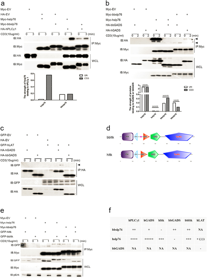
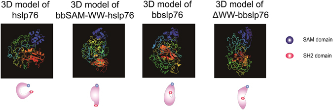
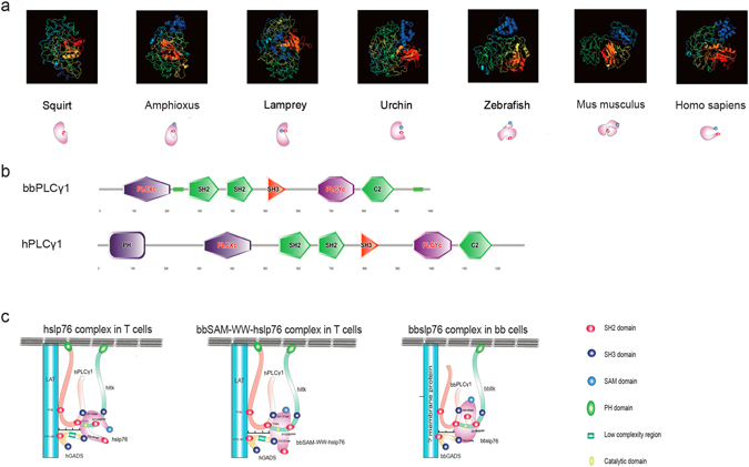
The well-defined mammalian slp76-signalosome is crucial for T-cell immune response, yet whether slp76-signalosome exists in invertebrates and how it evolved remain unknown. Here we investigated slp76-signalosome from an evolutionary perspective in amphioxus Branchiostoma belcheri (bb). We proved slp76-signalosome components bbslp76, bbGADS and bbItk are present in amphioxus and bbslp76 interacts with bbGADS and bbItk, but differences exist between the interaction manners within slp76-signalosome components of amphioxus and human (h). Specifically, bbslp76 has a unique WW-domain that blocked its association with hItk and decreased TCR-induced tyrosine-phosphorylation and NFAT-activation. Deletion of WW-domain shifted the constitutive association between bbslp76 and hPLCγ1 to a TCR-enhanced association. Among slp76-signalosome, the interaction between slp76 and PLCγ1 is the most conserved and the binding between Itk and slp76 evolved from constitutive to stimulation-regulated. Sequence alignment and 3D structural analysis of slp76-signalosome molecules from keystone species indicated slp76 evolved into a more unfolded and flexible adaptor due to lack of WW-domain and several low-complexity-regions (LCRs) while GADS turned into a larger protein by a LCR gain, thus preparing more space for nucleating the coevolving slp76-signalosome. Altogether, through deletion of WW-domain and manipulation of LCRs, slp76-signalosome evolves from a rigid and stimulation-insensitive to a more flexible and stimulation-responding complex.


Conflict of interest statement

The authors declare that they have no competing interests.

Figures

Figure 1
Figure 1
Sequence and phylogeny tree of bbslp76. (a) Nucleotide and amino acid sequence of bbslp76. Itk binding motif DYEPPP was marked in the red frame. (b) Protein structures of bbslp76 and hslp76 generated by SMART. (c) Sequences alignment of bbslp76 and hslp76. The color of Sky blue represents 50% consensus of amino acid and dark blue represents 100% consensus of amino acid. Itk binding motif DYEPPP motif was highlight with ******. (d) Phylogenetic tree of slp76. The neighbor-joining phylogenetic tree was constructed on the basis of 13 different slp76 sequences from GenBank, utilizing the sequence analysis tool MEGA 5.
Figure 2
Figure 2
Tissue expression patterns of bbslp76. (a) Relative expression levels of bbslp76 gene in various tissues of B. belcheri by qRT-PCR. 2-ΔΔCt values (normalized to endogenous control 18S) represented the expression of the bbslp76 gene in various tissues (muscle, intestine, gill, skin, ovum) of B. belcheri. Bars represent the average results of triplicate reactions for each tissue. Error bars represent the standard error of the mean (SEM) among three biological replicates. *, ** and *** indicate a significant expression level difference versus muscle as control at p < 0.05, p < 0.005 and p < 0.005, respectively. (b) bbslp76 polyclonal antibody titration (1:1000) in 293 T cells with exogenous expression of cMyc-bbslp76 and in amphioxus intestine lysate respectively. EV: Empty Vector. (c) and (d) Localization of bbslp76 in the gill (c) or intestine cells (d) of amphioxus. Amphioxus gill or intestine cells were bound to poly-L-Lysine-coated coverslips, fixed, and stained with anti-bbslp76 polyclonal antibody followed by a secondary fluorescent antibody (green). Nuclei were stained with DAPI (blue). Images were acquired using fluorescent microscopy at 400 times magnification. Bar = 2 μm.
Figure 3
Figure 3
bbslp76 inhibits TCR-induced NFAT activation. (a) bbslp76 inhibits the total tyrosine phosphorylation of Jurkat TAg cells after TCR ligation. Jurkat TAg cells were transfected with NC (negative control, scrambled small interfering RNA) and shslp76 (short hairpin RNA targeting hslp76 RNA specifically). shslp76-transfected cells were separately co-transfected with Myc-hslp76, Myc-bbslp76, and Myc-bbslp76 plus HA-bbGADS. After 48 hrs, cells stimulated with or without CD3 (10 μg/mL, 5 min) were subjected to western-blotting to analyze the tyrosine phosphorylation (pY) of T cells. (b) bbslp76 inhibits the phosphorylation of Erk (pErk) in Jurkat TAg cells after TCR ligation. Cells and lysates were prepared as in (a). (c) bbslp76 inhibits NFAT activity in T cells. Jurkat TAg cells were transfected with NC and sislp76 (small interfering RNA targeting hslp76 RNA specifically). NC and sislp76 cells were transfected with an NFAT reporter construct and a co-transfected Renilla luciferase and the indicated constructs. 48 hrs later, cells were untreated or stimulated with CD3 (3 μg/mL) for 8 hrs and lysed for the reporter gene assay. Luciferase activity was normalized using a co-transfected Renilla luciferase. The data represent the mean ± SD from triplicate determinations. *P < 0.05, **P < 0.005, ***P < 0.0005. (d) Protein structure of bbGADS and hGADS generated by SMART. And sequences alignment of bbGADS and hGADS. The color of Sky blue represents 50% consensus of amino acid and dark blue represents 100% consensus of amino acid. The experiments were repeated three times with very similar results.
Figure 4
Figure 4
bbslp76 interacts with TCR proximal signal proteins. (a) bbslp76 interacts with hPLCγ1 in T cells. Jurkat TAg cells were transfected with the indicated constructs. After 48 hrs, cells were stimulated with or without CD3 (10 μg/mL, 3 min); then, lysed and lysates were split into two parts, one part of the lysates was subjected to a Myc IP (immunoprecipitation), and the other part was used for WCL (whole cell lysis, 40 μL). Samples were studied by western blot, and probed with indicated antibodies. The binding affinity is normalized by IB HA to IP Myc. (b) The interaction of bbslp76 or hslp76 with bbGADS or hGADS in T cells. Cells and lysates were prepared as in (a). Samples were studied by western blot, and probed with indicated antibodies. The upper arrow indicates hGADS. The lower arrow indicates bbGADS. The binding affinity is normalized by IB HA to IP Myc. (c) bbGADS does not interact with hLAT in T cells. Cells and lysates were prepared as in (a). One part of the lysates was subjected to a HA IP. Samples were studied by western blot, and probed with indicated antibodies. The arrow indicates hLAT. (d) Protein structures of bbItk and hItk generated by SMART. (e) bbslp76 does not interact with hItk in T cells. Cells and Lysates were prepared as in (a). Samples were studied by western blot, and probed with indicated antibodies. (f) Summary of binding affinities between bbslp76 and TCR proximal signal proteins. NA: not available; The experiments were repeated three times with very similar results. The strength of protein-protein interaction was analyzed by software Quantity One.
Figure 5
Figure 5
Mapping bbslp76 inhibition domain. (a) Schematic structures of chimeric proteins. (b) Interactions of chimeric proteins with hItk. Jurkat TAg cells were transfected with the indicated constructs. After 48 hrs, cells were stimulated with or without CD3 (10 μg/mL, 1 min) and then lysed; lysates were split into two parts. One part of the lysates was subjected to a HA IP, and the other part was used for WCL (40 μL). Samples were studied by western blot, and probed with indicated antibodies. The binding affinity is normalized by IB GFP to IP HA. (c) bbslp76 inhibits NFAT activity in T cells. Jurkat TAg cells were transfected with NC and sislp76. NC and sislp76 cells were transfected with an NFAT reporter construct and a co-transfected Renilla luciferase and the indicated constructs. 48 hrs later, cells were untreated or stimulated with CD3 (3 μg/mL) for 8 hrs and lysed for reporter gene assay. Luciferase activity was normalized using a co-transfected Renilla luciferase. The data represent the mean ± SD from triplicate determinations. *P < 0.05, **P < 0.005, ***P < 0.0005. The experiments were repeated three times with very similar results. The strength of protein-protein interaction was analyzed by software Quantity One.
Figure 6
Figure 6
ΔWW-bbslp76 rescues total tyrosine phosphorylation but partially rescues NFAT activation after TCR ligation in T cells. (a) ΔWW-bbslp76 rescued total tyrosine phosphorylation in T cells. Jurkat TAg cells were transfected with the indicated constructs and cells and lysates were prepared as in Fig. 3(a). (b) bbslp76 partially rescued NFAT activity in T cells. Jurkat TAg cells were transfected with NC and shslp76. NC and shslp76 cells were transfected with an NFAT reporter construct and a co-transfected Renilla luciferase and the indicated constructs. 48 hrs later, cells were untreated or stimulated with CD3 (3 μg/mL) for 8 hrs and lysed for reporter gene assay. Luciferase activity was normalized using a co-transfected Renilla luciferase. The data represent the mean ± SD from triplicate determinations. *P < 0.05, **P < 0.005, ***P < 0.0005. (c) ΔWW-bbslp76 interacts with hItk. Jurkat TAg cells were transfected with the indicated constructs. After 48 hrs, cells were stimulated with or without CD3 (10 μg/mL, 3 min) and then lysed; lysates were split into two parts, one part of the lysates was subjected to a Myc IP, and the other part was used for WCL (40 μL). Samples were studied by western blot, and probed with indicated antibodies. (d) ΔWW-bbslp76 interacts with hPLCγ1. Jurkat TAg cells were transfected with the indicated constructs. After 48 hrs, cells were stimulated with or without CD3 (10 μg/mL, 1 min). Cells and lysates were prepared as in (c). Samples were studied by western blot, and probed with the indicated antibodies. The binding affinity is normalized by IB HA to IP Myc. The experiments were repeated three times with very similar results. The strength of protein-protein interaction was analyzed by software Quantity One.
Figure 7
Figure 7
3D model comparison of hslp76, bbSAM-WW-hslp76, bbslp76 and ΔWW-bbslp76. The 3D structures were predicted by Phyre2.
Figure 8
Figure 8
3D model comparison of slp76 from several keystone species. (a) 3D model comparison of slp76 from several keystone species. The 3D structures were predicted by Phyre2. (b) Protein structures of bbPLCγ1 and hPLCγ1generated by SMART. (c) A web of interactions connects hslp76 to the hPLCγ1, hItk, hLAT and hGADS. Predicted interactions of bbSAM-WW-hslp76 in T cells and bbslp76 in bb cells. Scales show the distance between LAT and slp76.

Similar articles

Cited by

References

    1. Brownlie RJ, Zamoyska R. T cell receptor signalling networks: branched, diversified and bounded. Nat Rev Immunol. 2013;13:257–269. doi: 10.1038/nri3403. - DOI - PubMed
    1. Malissen B, Bongrand P. Early T Cell Activation: Integrating Biochemical, Structural, and Biophysical Cues. Annual Review of Immunology. 2015;33:539–561. doi: 10.1146/annurev-immunol-032414-112158. - DOI - PubMed
    1. Wang H, et al. ZAP-70: an essential kinase in T-cell signaling. Cold Spring Harb Perspect Biol. 2010;2:a002279–a002279. doi: 10.1101/cshperspect.a002279. - DOI - PMC - PubMed
    1. Balagopalan L, Coussens NP, Sherman E, Samelson LE, Sommers CL. The LAT story: a tale of cooperativity, coordination, and choreography. Cold Spring Harb Perspect Biol. 2010;2:a005512–a005512. doi: 10.1101/cshperspect.a005512. - DOI - PMC - PubMed
    1. Jordan MS, Koretzky GA. Coordination of receptor signaling in multiple hematopoietic cell lineages by the adaptor protein SLP-76. Cold Spring Harb Perspect Biol. 2010;2:a002501–a002501. doi: 10.1101/cshperspect.a002501. - DOI - PMC - PubMed

Publication types

MeSH terms

Substances

LinkOut - more resources