. 2017 Apr 20:8:642.
doi: 10.3389/fmicb.2017.00642. eCollection 2017.

FnrL and Three Dnr Regulators Are Used for the Metabolic Adaptation to Low Oxygen Tension in Dinoroseobacter shibae

Affiliations

FnrL and Three Dnr Regulators Are Used for the Metabolic Adaptation to Low Oxygen Tension in Dinoroseobacter shibae

Matthias Ebert et al. Front Microbiol. .

Abstract

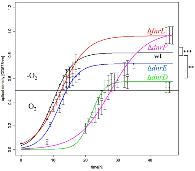
The heterotrophic marine bacterium Dinoroseobacter shibae utilizes aerobic respiration and anaerobic denitrification supplemented with aerobic anoxygenic photosynthesis for energy generation. The aerobic to anaerobic transition is controlled by four Fnr/Crp family regulators in a unique cascade-type regulatory network. FnrL is utilizing an oxygen-sensitive Fe-S cluster for oxygen sensing. Active FnrL is inducing most operons encoding the denitrification machinery and the corresponding heme biosynthesis. Activation of gene expression of the high oxygen affinity cbb3-type and repression of the low affinity aa3-type cytochrome c oxidase is mediated by FnrL. Five regulator genes including dnrE and dnrF are directly controlled by FnrL. Multiple genes of the universal stress protein (USP) and cold shock response are further FnrL targets. DnrD, most likely sensing NO via a heme cofactor, co-induces genes of denitrification, heme biosynthesis, and the regulator genes dnrE and dnrF. DnrE is controlling genes for a putative Na+/H+ antiporter, indicating a potential role of a Na+ gradient under anaerobic conditions. The formation of the electron donating primary dehydrogenases is coordinated by FnrL and DnrE. Many plasmid encoded genes were DnrE regulated. DnrF is controlling directly two regulator genes including the Fe-S cluster biosynthesis regulator iscR, genes of the electron transport chain and the glutathione metabolism. The genes for nitrate reductase and CO dehydrogenase are repressed by DnrD and DnrF. Both regulators in concert with FnrL are inducing the photosynthesis genes. One of the major denitrification operon control regions, the intergenic region between nirS and nosR2, contains one Fnr/Dnr binding site. Using regulator gene mutant strains, lacZ-reporter gene fusions in combination with promoter mutagenesis, the function of the single Fnr/Dnr binding site for FnrL-, DnrD-, and partly DnrF-dependent nirS and nosR2 transcriptional activation was shown. Overall, the unique regulatory network of the marine bacterium D. shibae for the transition from aerobic to anaerobic growth composed of four Crp/Fnr family regulators was elucidated.

Keywords: Crp/Fnr regulator; Dinoroseobacter shibae; Dnr; FnrL; anaerobic energy metabolism; denitrification; oxygen-dependent gene expression; regulation.


Figures

Figure 1
Figure 1
Phylogenetic affiliations of the D. shibae Crp/Fnr-like regulators. (A) The unrooted neighbor joining trees containing D. shibae Crp/Fnr like regulators affiliated to the Crp/Fnr superfamily are shown. (B) FnrN subfamily of Fnr like regulators containing FnrL of D. shibae. (C) Dnr subgroup of Crp/Fnr like regulators containing three different Dnr regulators of D. shibae. The members of the tree are identified by the name of the host bacterium and the NCBI sequence identification number (accession number).
Figure 2
Figure 2
Alignment of the N-terminal regulatory domains of FnrL like regulators. Cysteine residues which were known to be essential for activity in P. denitrificans are marked in red (Hutchings et al., 2002). Those cysteine residues which were additionally found conserved exclusively in the Roseobacter group are indicated in blue. The alignment comprises the amino acid sequences of FnrL proteins from Dinoroseobacter shibae DFL12T (WP_012177338.1), Paracoccus denitrificans (WP_041529894.1), Rhodobacter sphaeroides (WP_002720880.1), Roseobacter litoralis (WP_044025718.1), Roseobacter denitrificans (WP_044032974.1), Pheobacter gallaeciensis (WP_014881157.1), Phaeobacter. inhibens (WP_040174583.1), Roseibacterium elongatum (WP_025310970.1), Ruegeria pomeroyi (WP_011049211.1), Leisingera methylohalidivorans (WP_024088845.1), Ruegeria sp. (WP_039983362.1), Jannaschia sp. (WP_011456972.1), Paracoccus aminophilus (WP_020949853.1), Octadecabacter antarcticus (WP_015498258.1), Octadecabacter arcticus 238 (WP_015493613.1), Ketoglonicigernium vulgare (WP_013383280.1). In addition, Fnr from Escherichia coli (WP_000611911.1) and Anr from Pseudomonas putida (WP_010954166.1) were included. Symbols indicates an entirely conserved column (*), a column comprising amino acids of same size and hydropathy (:) and a column comprising amino acids of similar size or evolutionary preserved hydropathy (.) (Notredame et al., 2000).
Figure 3
Figure 3
FnrL contains an oxygen labile Fe-S cluster (A) Reconstitution of the D. shibae DFL12T FnrL Fe-S clusters. The spectrum of 40 μM FnrL (black line) and 20 μM Fe-S cluster reconstituted FnrL (red line) are shown. Samples were normalized by setting absorption at 280 nm to 1. (B) Oxygen lability of the FnrL Fe-S cluster. The exposure of Fe-S cluster reconstituted FnrL to air resulted in a decrease of absorption at 420 nm over time [0 min (red line), 60 min (blue line), 120 min (magenta line), and 240 min (green line)].
Figure 4
Figure 4
Growth behavior of D. shibae Dfl12T and oxygen regulatory mutants during an aerobic to anaerobic shift. D. shibae DFL12T wildtype strain (black line), DS001(ΔfnrL) (red line), DS002(ΔdnrD) (green line), DS003(ΔdnrE) (blue line) and DS004(ΔdnrF) (magenta line) mutant strains were grown under aerobic conditions in artificial see water medium supplemented with 16.9 mM succinate. After reaching of OD578 nm of 0.5 cells were shifted to anaerobic growth conditions (black horizontal line) and 25 mM sodium nitrate was added. The optical density was measured in three independent replicates and error bars represent the standard deviation. P-values were determined by using ANOVA without a Post-hoc-test using the wild type values as reference. The symbols indicate P-values smaller or equal to 0 (***) or 0.001 (**).
Figure 5
Figure 5
Regulation of the denitrification operons by FnrL, DnrD, DnrE, and DnrF. (A) Heat map representation of denitrification gene expression patterns of mutant strains DS001(ΔfnrL), DS002(ΔdnrD), DS003(ΔdnrE), and DS004(ΔdnrF) compared to D. shibae DFL12T wild type strain grown under anaerobic conditions. The colored bars represent the expression level in log2 scale. Green indicates a relatively low expression level in the mutant strain which indicates activation by the regulator; red indicates relatively high expression levels in the mutant strain compared to the wild type indicating repression. (B) Denitrification operons with corresponding RNA sequencing data based transcriptional start sites and transcript quantification under aerobic (blue line) and anaerobic conditions (red line) of the wild type D. shibae DFL12T. Black horizontal arrows indicate open reading frames and the direction of transcription. Additionally, binding sites of FnrL, DnrD, DnrE, and DnrF are indicated by flags. If the corresponding binding site was located within a promoter sequence, the corresponding distance to the transcriptional start was given. Green boxes indicate an activation by the given regulator, red boxes indicate a repression. a1, apbE1; c1, cycA1; hyp, hypothetical gene; dD, dnrD; hA, hemA3; dE, dnrE.
Figure 6
Figure 6
Functional investigation of the nirSnosR2 intergenic promoter region using nirS-lacZ and nosR2-lacZ reporter gene fusions. D. shibae DFL12T wild type (WT), DS001(ΔfnrL) (ΔfnrL), DS002(ΔdnrD) (ΔdnrD), DS003(ΔdnrE) (ΔdnrE), and DS004(ΔdnrF) (ΔdnrF) mutant strains carrying the nirS-lacZ and nosR2-lacZ reporter gene fusions were grown under oxygen-limited conditions, and β-galactosidase activity was measured. Error bars represent the observed standard deviation. P-values were determined using ANOVA and the Tukey-test (Tukey, 1949). The symbol *** indicate P-values smaller or equal to 0. The palindromic sequence of the Fnr/Dnr binding site and its position with respect to the transcriptional start site of nirS and nosR2 are given.
Figure 7
Figure 7
Functional importance of the Fnr/Dnr binding site in the intergenic region of nirS and nosR2. D. shibae wild type strains DFL12T carrying the nirS-lacZ and nirS(mu)-lacZ reporter gene fusions were grown under aerobic and anaerobic growth conditions and β-galactosidase activities were measured in three independent replicates. The nirS-lacZ reporter gene fusion is carrying the palindromic sequence 5′-TTAAC-N4-GTCAA-3′ of the Fnr/Dnr binding site. The binding sequence was mutated to 5′-GCAAC-N4-GTCGC-3′ in the nirS(mu)-lacZ reporter gene fusion. Error bars represent the observed standard deviation. P-values were determined using ANOVA and the Tukey-test (Tukey, 1949). The symbol *** indicate P-values smaller or equal to 0.
Figure 8
Figure 8
Model for the regulation of the energy metabolism of D. shibae by FnrL, Dnr D, DnrE and DnrF during the aerobic to anaerobic transition. DH, dehydrogenase; Cyt, cytochrome; bchl, bacteriochlorophyll; Qdand Qb oxidized and reduced form of reaction center quinone, respectively; Cox, cytochrome oxidase; DMSO, dimethyl sulfoxide; Red, reductase. Figure was adapted from Wagner-Döbler et al. (2010).
Figure 9
Figure 9
Venn diagram of the overlapping regulons of Fnr, DnrD, DnrE and DnrF derived from transcriptional profiling experiments. Numbers for differentially expressed genes regulated by indicated regulators determined by transcriptome analyses using mutants of the corresponding regulator genes compared for aerobic vs. anaerobic growth are given.
Figure 10
Figure 10
Cascade type of regulatory network for the control of the aerobic-anaerobic transition by the Crp/Fnr like regulators in D. shibae DFL12T. FnrL sensing oxygen via a bound oxygen-labile Fe-S cluster is the main oxygen regulator. DnrD most likely sensing NO via a bound heme cofactor is the main regulator for denitrification. the master regulators of the aerobic-anaerobic transition. They are inducing/repressing multiple transcriptional units of indicated processes. Moreover, expression of dnrE and dnrF, which in turn are controlling their own regulons, are modulating the activity of FnrL and DnrD. Interestingly, DnrE is mainly controlling plasmid-encoded genes.

References

    1. Arai H. (2003). Transcriptional regulation of the nos genes for nitrous oxide reductase in Pseudomonas aeruginosa. Microbiology 149, 29–36. 10.1099/mic.0.25936-0 - DOI - PubMed
    1. Arai H., Igarashi Y., Kodama T. (1995). Expression of the nir and nor genes for denitrification of Pseudomonas aeruginosa requires a novel CRP/ FNR-related transcriptional regulator, DNR, in addition to ANR. FEBS Lett. 371, 73–76. 10.1016/0014-5793(95)00885-D - DOI - PubMed
    1. Becher A., Schweizer H. P. (2000). Integration-proficient Pseudomonas aeruginosa vectors for isolation of single-copy chromosomal lacZ and lux gene fusions. Biotechniques 29, 948–952. - PubMed
    1. Biebl H., Allgaier M., Tindall B. J., Koblizek M., Lünsdorf H., Pukall R. (2005). Dinoroseobacter shibae gen. nov., sp. nov., a new aerobic phototrophic bacterium isolated from dinoflagellates. Int. J. Syst. Evol. Microbiol. 55, 1089–1096. 10.1099/ijs.0.63511-0 - DOI - PubMed
    1. Blanka A., Schulz S., Eckweiler D., Franke R., Bielecka A., Nicolai T., et al. . (2014). Identification of the alternative sigma factor SigX regulon and its implications for Pseudomonas aeruginosa pathogenicity. J. Bacteriol. 196, 345–356. 10.1128/JB.01034-13 - DOI - PMC - PubMed