

Posterior reversible encephalopathy syndrome(PRES)
- PMID: 28473920
- PMCID: PMC5410886
- DOI: 10.1093/omcr/omx011
Posterior reversible encephalopathy syndrome(PRES)
Abstract
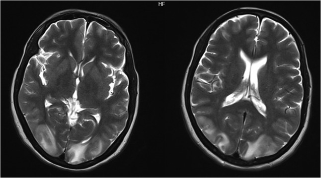
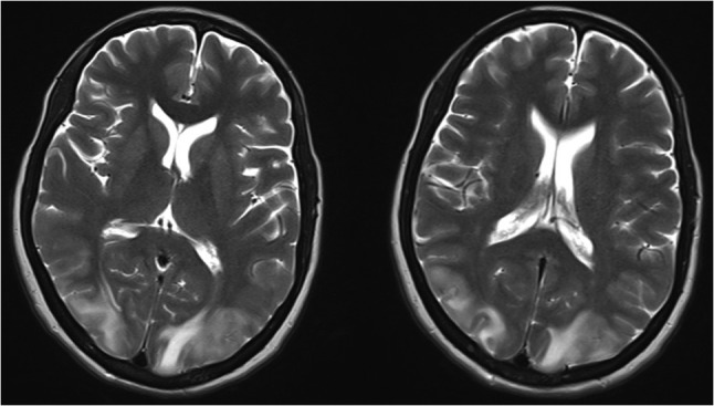
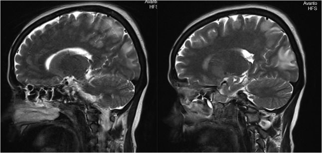
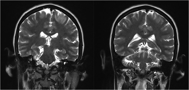
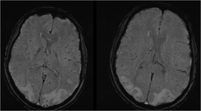
Posterior reversible encephalopathy syndrome (PRES) is a clinico-radiological syndrome characterized by a headache, seizures, altered mental status and visual loss and characterized by white matter vasogenic edema affecting the posterior occipital and parietal lobes of the brain predominantly. This clinical syndrome is increasingly recognized due to improvement and availability of brain imaging specifically magnetic resonance imaging (MRI). A 35-year-old female with the history of unsafe abortion and massive blood transfusion 10 days ago was brought to the emergency room with three episodes of generalized tonic-clonic seizures, urinary incontinence and altered sensorium since 3 hours. MRI brain showed bilateral occipital, parietal, frontal cortex and subcortical white matter T2/Fluid-attenuated inversion recovery hyperintensities, suggestive of PRES. The patient improved after management with intravenous fluids, antibiotics, antiepileptics and monitoring of blood pressure. If recognized and treated early, the clinical syndrome commonly resolves within a week. PRES can be a major problem in rapid and massive blood transfusion. A high index of suspicion and prompt treatment can reduce morbidity, mortality and pave the path for early recovery.
Figures
References
-
- McKinney AM, Short J, Truwit CL, McKinney ZJ, Kozak OS, SantaCruz KS, et al. Posterior reversible encephalopathy syndrome: incidence of atypical regions of involvement and imaging findings. Am J Roentgenol 2007;189:904–12. - PubMed
-
- Hinchey J, Chaves C, Appignani B, Breen J, Pao L, Wang A, et al. A reversible posterior leukoencephalopathy syndrome. New Engl J Med 1996. Feb 22;334:494–500. - PubMed
-
- Schwartz RB, Jones KM, Kalina P, Bajakian RL, Mantello MT, Garada B, et al. Hypertensive encephalopathy: findings on CT, MR imaging, and SPECT imaging in 14 cases. AJR Am J Roentgenol 1992;159:379–83. - PubMed
Publication types
LinkOut - more resources
Full Text Sources
Other Literature Sources
