

Amide proton transfer CEST of the cervical spinal cord in multiple sclerosis patients at 3T
- PMID: 28474409
- PMCID: PMC5777527
- DOI: 10.1002/mrm.26736
Amide proton transfer CEST of the cervical spinal cord in multiple sclerosis patients at 3T
Abstract
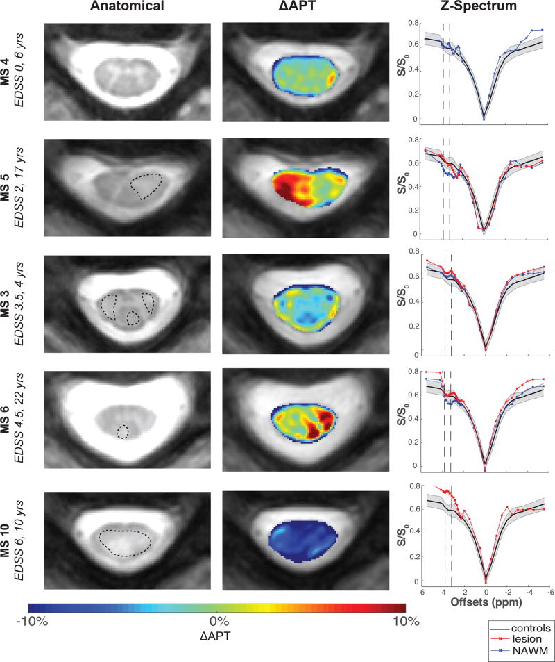
Purpose: The ability to evaluate pathological changes in the spinal cord in multiple sclerosis (MS) is limited because T1 - and T2 -w MRI imaging are not sensitive to biochemical changes in vivo. Amide proton transfer (APT) chemical exchange saturation transfer (CEST) can indirectly detect amide protons associated with proteins and peptides, potentially providing more pathological specificity. Here, we implement APT CEST in the cervical spinal cord of healthy and MS cohorts at 3T.
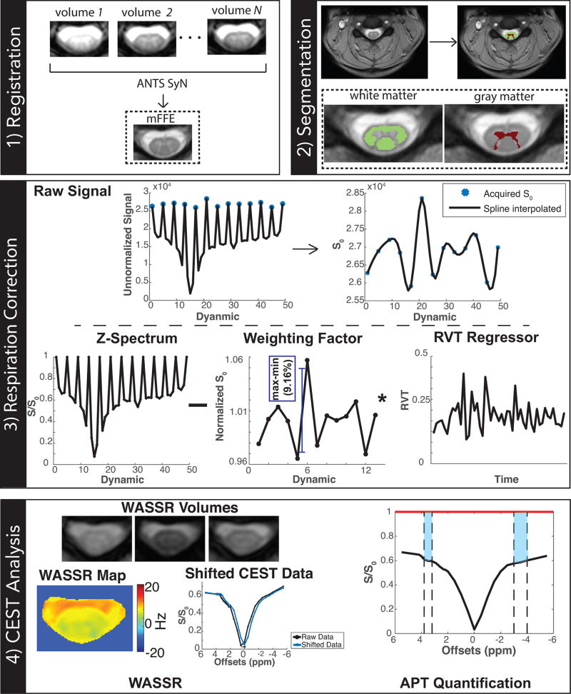
Methods: APT CEST of the cervical spinal cord was obtained in a cohort of 10 controls and 10 MS patients using a novel respiratory correction methodology. APT was quantified using two methods: 1) APTw , based off the conventional magnetization transfer ratio asymmetry, and 2) ΔAPT, a spatial characterization of APT changes in MS patients relative to the controls.
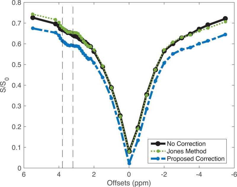
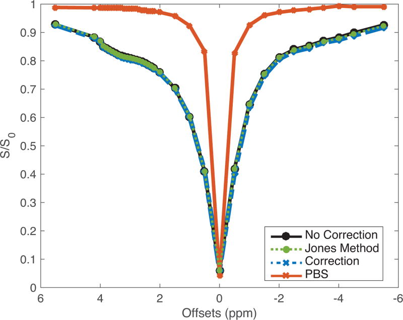
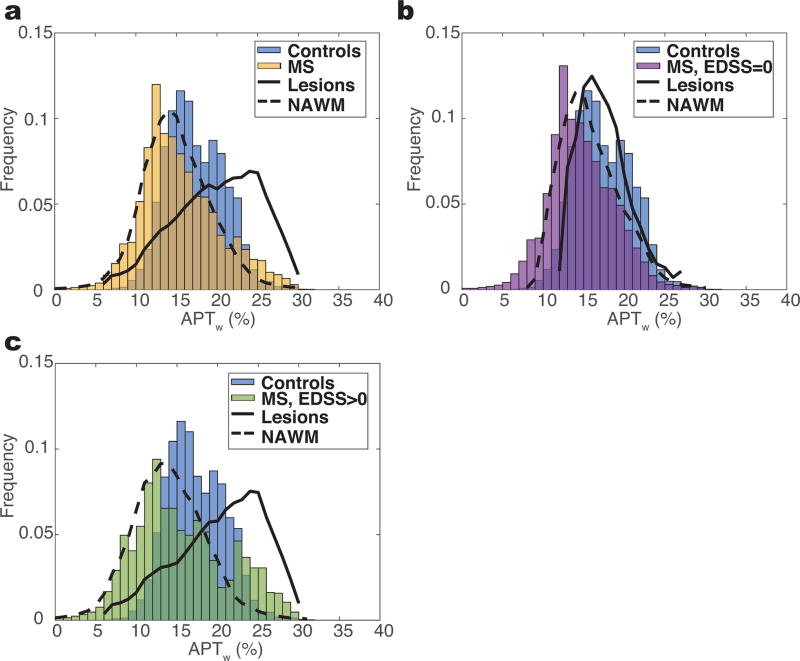
Results: Respiratory correction yielded highly reproducible z-spectra in white matter (intraclass correlation coefficient = 0.82). APTw signals in normal-appearing white matter (NAWM) of MS patients were significantly different from healthy controls (P = 0.04), whereas ΔAPT of MS patients highlighted large APT differences in NAWM.
Conclusion: Respiration correction in the spinal cord is necessary to accurately quantify APT CEST, which can provide unique biochemical information regarding disease processes within the spinal cord. Magn Reson Med 79:806-814, 2018. © 2017 International Society for Magnetic Resonance in Medicine.
Keywords: CEST; amide proton transfer; multiple sclerosis; normal-appearing white matter; spinal cord.
© 2017 International Society for Magnetic Resonance in Medicine.
Figures
References
-
- Bot JC, Barkhof F, Polman CH, Lycklama a Nijeholt GJ, de Groot V, Bergers E, Ader HJ, Castelijns JA. Spinal cord abnormalities in recently diagnosed MS patients: added value of spinal MRI examination. Neurology. 2004;62(2):226–233. - PubMed
-
- Kearney H, Miller DH, Ciccarelli O. Spinal cord MRI in multiple sclerosis-diagnostic, prognostic and clinical value. Nat Rev Neurol. 2015;11(6):327–338. - PubMed
-
- Bergers E, Bot JC, De Groot CJ, Polman CH, Lycklama a Nijeholt GJ, Castelijns JA, van der Valk P, Barkhof F. Axonal damage in the spinal cord of MS patients occurs largely independent of T2 MRI lesions. Neurology. 2002;59(11):1766–1771. - PubMed
-
- Zhou J, Payen J-F, Wilson DA, Traystman RJ, van Zijl PCM. Using the amide proton signals of intracellular proteins and peptides to detect pH effects in MRI. Nat Med. 2003;9(8):1085–1090. - PubMed
Publication types
MeSH terms
Substances
Grants and funding
LinkOut - more resources
Full Text Sources
Other Literature Sources
Medical
