. 2017 Jun;5(2):109-123.
doi: 10.1002/iid3.146. Epub 2017 Feb 20.

Innate CD8αα+ lymphocytes enhance anti-CD40 antibody-mediated colitis in mice

Affiliations

Innate CD8αα+ lymphocytes enhance anti-CD40 antibody-mediated colitis in mice

Aaram A Kumar et al. Immun Inflamm Dis. 2017 Jun.

Abstract

Introduction: Immune responses in the intestines require tight regulation to avoid uncontrolled inflammation. We previously described an innate lymphocyte population in the intestinal epithelium (referred to as innate CD8αα+ , or iCD8α cells) that can protect against gastrointestinal infections such as those mediated by Citrobacter rodentium.

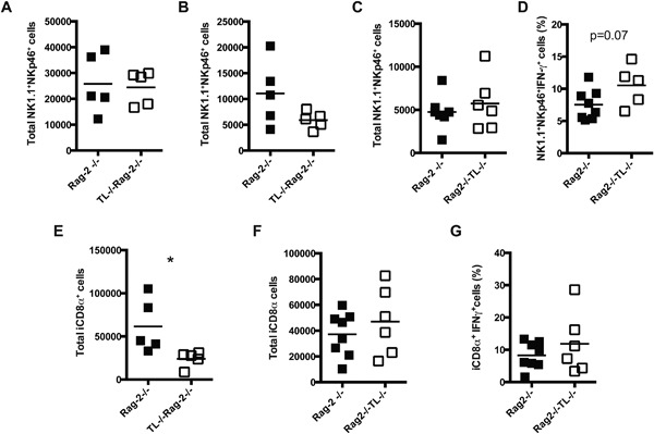
Methods: Here, we have evaluated the potential contribution of these cells to intestinal inflammation by analyzing inflammation development in mice with decreased numbers of iCD8α cells. We also determined the potential of iCD8α cells to secrete granzymes and their potential role during inflammatory processes.

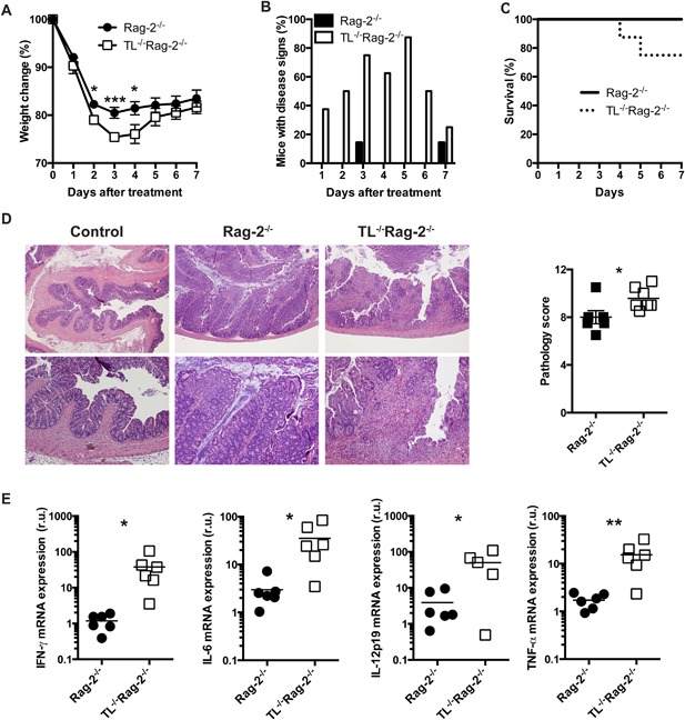
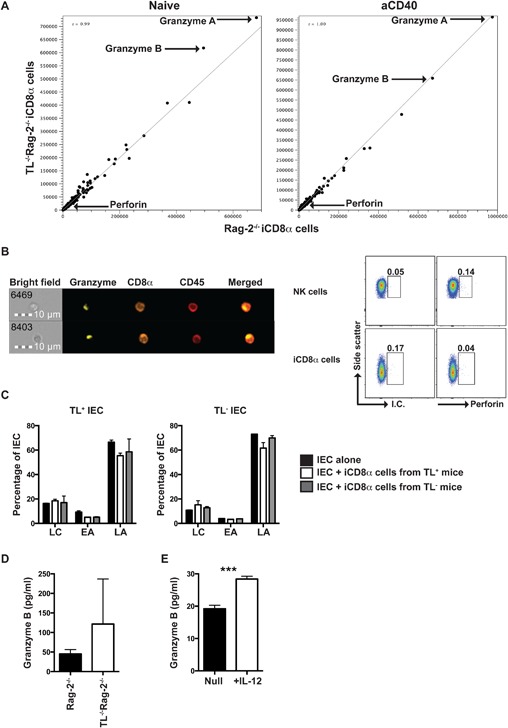
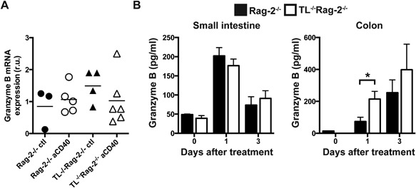
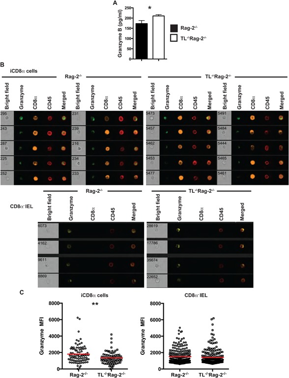
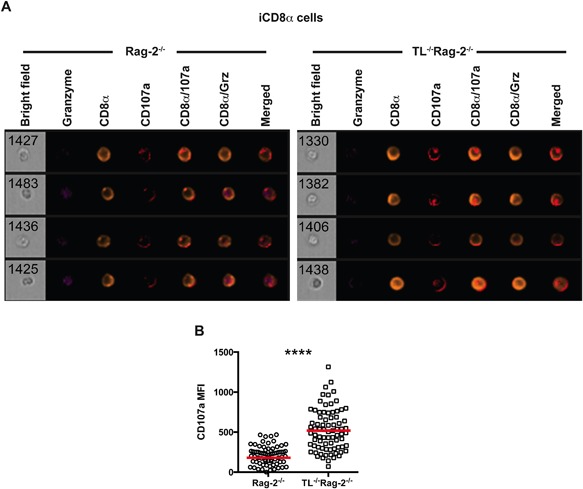
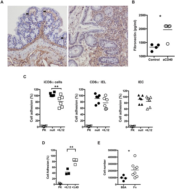
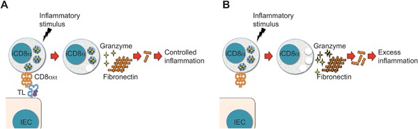
Results: We found that iCD8α cells play a pro-inflammatory role in the development of disease in a colitis model induced by anti-CD40 antibodies. We further found that the effects of iCD8α cells correlated with their capacity to secrete granzymes. We also observed that the pro-inflammatory properties of iCD8α cells were controlled by interactions of CD8αα homodimers on these cells with the thymus leukemia antigen expressed by intestinal epithelial cells.

Conclusions: Our findings suggest that iCD8α cells modulate inflammatory responses in the intestinal epithelium, and that dysregulation of iCD8α cells effector functions may enhance disease. We propose that one of the mechanism by which iCD8α cells enhance inflammation is by the secretion of granzymes, which may promote recruitment of infiltrating cells into the epithelium.

Keywords: Granzyme; innate CD8ɑɑ cells; intestinal inflammation; intraepithelial lymphocytes.


Figures

Figure 1
Figure 1
TL‐deficiency renders Rag‐2−/− mice more susceptible to anti‐CD40‐induced colitis. Rag‐2−/− and TL−/−Rag‐2−/− mice were treated with 150 μg/mouse of anti‐CD40 antibodies and were monitored daily for (A) weight change, (B) signs of disease such as rectal bleeding, diarrhea and scruffiness, and (C) survival. (D) Colon pathology was determined seven days after treatment as described in the Materials and methods section. Top images, 40× magnification; bottom images, 100× magnification. The graph summarizes the pathology scores. (E) Total colon cytokine mRNA expression was measured by real‐time PCR. Colons from naïve mice were used as reference. The data shown are representative of more than three similar experiments (n = 6/experiment). Error bars indicate standard deviation. Student's t test was performed. *P < 0.05; **P < 0.01; ***P < 0.005.
Figure 2
Figure 2
iCD8α cells expand in TL‐deficient mice after anti‐CD40 treatment. Total NK1.1+NKp46+ IEL numbers in (A) naïve Rag‐2−/− and TL−/−Rag‐2−/− mice mice, or animals (B) 1, and (C) 2 days post anti‐CD40 antibodies treatment. (D) Frequencies of NK1.1+NKp46+ IFN‐γ+ IEL from Rag‐2−/− and TL−/−Rag‐2−/− mice 2 days post anti‐CD40 treatment. Total iCD8α cell numbers in Rag‐2−/− and TL−/−Rag‐2−/− mice after (E) 1 and (F) 2 days treatment with anti‐CD40 antibodies. (G) Frequencies of IFN‐γ+ iCD8α cells in Rag‐2−/− and TL−/−Rag‐2−/− mice 2 days post‐treatment. The data shown are representative of two independent experiments. Each symbol indicates an individual mouse. Student's t test was performed. *P < 0.05.
Figure 3
Figure 3
iCD8α cells express large quantities of granzymes. (A) RNA‐seq analysis of iCD8α cells derived from naïve (left) or 1‐day post anti‐CD40 treatment (right) Rag‐2−/− and TL−/−Rag‐2−/−. Data represent cells derived from at least 10 mice from each group. (B) Granzyme intracellular localization in iCD8α cells derived from naïve Rag‐2−/− mice. The number on the bright field quadrant identifies the acquired cell analyzed. Data show two representative cells of at least 50 cells analyzed with similar results (left image). Spleen NK cells or iCD8α cells were stained for intracellular perforin (right dot plots). (C) IEC derived from TL‐competent or TL‐deficient mice were cultured in the presence or absence of iCD8α cells derived from TL‐competent or TL‐deficient mice for four hours. At the end of the incubation period, IEC were stained for annexin V surface expression. LC, live cells; EA, early apoptosis; LA, late apoptosis. (D) Granzyme secretion by iCD8α cells derived from naïve Rag‐2−/− and TL−/−Rag‐2−/− mice. The data shown are representative of two independent experiments. Cells were isolated and pooled from at least 10 mice. (E) Granzyme secretion by IL‐12‐treated or untreated iCD8α cells. The data shown are representative of two independent experiments. Cells were isolated and pooled from at least 10 mice. Error bars indicate standard deviation. Student's t test was performed. ***P < 0.005.
Figure 4
Figure 4
Granzyme levels in the intestines of anti‐CD40‐treated mice. (A) Total granzyme B mRNA expression in the colons of Rag‐2−/− and TL−/−Rag‐2 1 day after treatment with anti‐CD40 antibodies or PBS (ctl). Each symbol represents an individual mouse. (B) Granzyme B levels in the small intestine (left) and colon (right) during the indicated time points after anti‐CD40 treatment. The data shown are representative of 2 independent experiments, (n = 5 or 6/experiment). Error bars indicate standard deviation. Student's t test was performed. *P < 0.05.
Figure 5
Figure 5
iCD8α cells from TL‐deficient mice secrete increased levels of granzymes. (A) iCD8α cells from anti‐CD40‐treated Rag‐2−/− and TL−/−Rag‐2−/− mice (2 days post‐treatment) were incubated overnight and granzyme concentration was determined by ELISA. The data shown are representative of at least two independent experiments, with 5–10 mice per experimental group. Error bars indicate standard deviation. (B) Total IEL from anti‐CD40‐treated Rag‐2−/− and TL−/−Rag‐2−/− mice (2 days post‐treatment) were analyzed for intracellular granzyme expression. Top images represent the analysis of iCD8α cells, and the bottom images represent CD45+CD8α IEL. (C) The graphs at the bottom represents a summary of at least 80 iCD8α or CD45+CD8α IEL cells analyzed for intracellular granzyme intensity from the indicated mice. The data shown are representative of at least three independent experiments and show pooled IEL from at least five mice. Each symbol represents an individual iCD8α cell. Colored bars indicate mean. Student's t test was performed. *P < 0.05; **P < 0.01.
Figure 6
Figure 6
iCD8α cells from TL‐deficient mice express increased surface levels of CD107a. (A) iCD8α cells from anti‐CD40‐treated Rag‐2−/− (left) and TL−/−Rag‐2−/− mice (right) were analyzed 2 days post‐treatment for CD107a surface expression. Four representative cells for each group are shown. (B) After acquisition, images were analyzed for CD107a mean fluorescence intensity. The data shown are representative of at least two independent experiments and show pooled IEL from at least three mice. Each symbol represents an individual iCD8α cell. Colored bars indicate mean. Student's t test was performed ****P < 0.001.
Figure 7
Figure 7
iCD8α cell‐derived granzymes digest fibronectin and promote cell infiltration. (A) Colon fibronectin staining (brown) from naïve (left) or anti‐CD40 treated (right) Rag‐2−/− mice. Arrows indicate areas of close proximity between the epithelium and fibronectin. Magnification, 40×. (B) Soluble fibronectin in total colon cultures of naïve and anti‐CD40‐treated Rag‐2−/− mice (2 days post‐treatment). Each symbol represents an individual mouse. (C) Specific B16 melanoma cell adhesion onto fibronectin‐coated plates treated with supernatants from cultured iCD8α cells (left), CD8α IEL (middle) and IEC (right) in the presence or absence of IL‐12. PK, proteinase K‐treated wells. (D) 3,4‐dichloroisocoumurin (1 mM) was included in IL‐12‐stimulated iCD8α cell supernatants. (E) Mice were injected i.p. with supernatant of fibronectin or BSA digests. Four hours later, leukocyte recruitment was evaluated by determining total cellularity in the peritoneum cavity. Each symbol represents an individual sample/mouse. Data represents the combination of two independent experiments. Student's t test was performed *P < 0.05; **P < 0.01.
Figure 8
Figure 8
Proposed model for iCD8α cell function in a TL‐competent or −deficient environment. (A) During inflammatory stimuli the TL‐CD8αα interaction regulates the effector function of iCD8α cells. In this scenario, relatively low levels of granzymes are secreted into the epithelium environment, resulting in controlled inflammation. (B) However, when TL is absent, iCD8α cells become dysregulated and inflammatory stimuli may induce these cells to secrete comparatively larger quantities of granzymes, which in turn enhances inflammation.

Similar articles

Cited by

References

    1. Cheroutre, H. , Lambolez F., and Mucida D.. 2011. The light and dark sides of intestinal intraepithelial lymphocytes. Nat. Rev. Immunol. 11:445–456. - PMC - PubMed
    1. Olivares‐Villagomez, D. , and Van Kaer L.. 2010. TL and CD8alphaalpha: enigmatic partners in mucosal immunity. Immunol. Lett. 134:1–6. - PMC - PubMed
    1. Cheroutre, H. , and Lambolez F.. 2008. Doubting the TCR coreceptor function of CD8alphaalpha. Immunity 28:149–159. - PubMed
    1. Chen, Y. , Chou K., Fuchs E., Havran W. L., and Boismenu R.. 2002. Protection of the intestinal mucosa by intraepithelial gamma delta T cells. Proc. Natl. Acad. Sci. USA 99:14338–14343. - PMC - PubMed
    1. Das, G. , Augustine M. M., Das J., Bottomly K., Ray P., and Ray A.. 2003. An important regulatory role for CD4 + CD8 alpha alpha T cells in the intestinal epithelial layer in the prevention of inflammatory bowel disease. Proc. Natl. Acad. Sci. USA 100:5324–5329. PMC1535703. - PMC - PubMed

Publication types